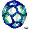
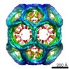
Journal: Nat Struct Mol Biol / Year: 2016 Title: Clathrin-coat disassembly illuminates the mechanisms of Hsp70 force generation. Authors: Rui Sousa / Hsien-Shun Liao / Jorge Cuéllar / Suping Jin / José M Valpuesta / Albert J Jin / Eileen M Lafer / Abstract: Hsp70s use ATP hydrolysis to disrupt protein-protein associations and to move macromolecules. One example is the Hsc70- mediated disassembly of the clathrin coats that form on vesicles during ...Hsp70s use ATP hydrolysis to disrupt protein-protein associations and to move macromolecules. One example is the Hsc70- mediated disassembly of the clathrin coats that form on vesicles during endocytosis. Here, we exploited the exceptional features of these coats to test three models-Brownian ratchet, power-stroke and entropic pulling-proposed to explain how Hsp70s transform their substrates. Our data rule out the ratchet and power-stroke models and instead support a collision-pressure mechanism whereby collisions between clathrin-coat walls and Hsc70s drive coats apart. Collision pressure is the complement to the pulling force described in the entropic pulling model. We also found that self-association augments collision pressure, thereby allowing disassembly of clathrin lattices that have been predicted to be resistant to disassembly. These results illuminate how Hsp70s generate the forces that transform their substrates.
History
Deposition
Jun 20, 2016
-
Header (metadata) release
Jul 27, 2016
-
Map release
Jul 27, 2016
-
Update
Aug 3, 2016
-
Current status
Aug 3, 2016
Processing site: PDBe / Status: Released
-
Structure visualization
Movie
Surface view with section colored by density value
Organism: Escherichia coli (E. coli) / Recombinant strain: BL21
Sequence
UniProtKB: Clathrin coat assembly protein AP180
-
Macromolecule #2: Bovine Auxilin residues 547-910
Macromolecule
Name: Bovine Auxilin residues 547-910 / type: protein_or_peptide / ID: 2 / Number of copies: 108 / Recombinant expression: Yes
Source (natural)
Organism: Bos taurus (cattle) / synonym: Cow
Molecular weight
Experimental: 38.935 KDa
Recombinant expression
Organism: Escherichia coli (E. coli) / Recombinant strain: BL21
Sequence
UniProtKB: Auxilin
-
Macromolecule #3: Rat Clathrin Light Chain A1: residues 1-248
Macromolecule
Name: Rat Clathrin Light Chain A1: residues 1-248 / type: protein_or_peptide / ID: 3 / Name.synonym: CLC, CLCA, CLCA1 / Number of copies: 108 / Recombinant expression: Yes
Source (natural)
Organism: Rattus norvegicus (Norway rat) / synonym: Rat
Molecular weight
Experimental: 26.979 KDa
Recombinant expression
Organism: Escherichia coli (E. coli) / Recombinant strain: BL21
Sequence
UniProtKB: Clathrin light chain A
-
Macromolecule #4: Rat Clathrin Heavy Chain 1: residues 1-1675
Macromolecule
Name: Rat Clathrin Heavy Chain 1: residues 1-1675 / type: protein_or_peptide / ID: 4 / Name.synonym: CHC, CHC1 / Details: His tag at the N-terminus / Number of copies: 108 / Recombinant expression: Yes
Source (natural)
Organism: Rattus norvegicus (Norway rat) / synonym: Rat
Molecular weight
Experimental: 196.358 KDa
Recombinant expression
Organism: Escherichia coli (E. coli) / Recombinant strain: BL21
Sequence
UniProtKB: Clathrin heavy chain 1
-
Experimental details
-
Structure determination
Method
cryo EM
Processing
single particle reconstruction
Aggregation state
particle
-
Sample preparation
Buffer
pH: 6 Details: 20 mM MES, 2 mM MgCl2, 25 mM KCl, 10 mM (NH4)2SO4 and 2 mM DTT
Grid
Details: Cu/Rh 300 mesh Quantifoil R 1.2/1.3 um grids with thin carbon support
Vitrification
Cryogen name: ETHANE / Chamber humidity: 90 % / Chamber temperature: 98 K / Instrument: LEICA EM CPC / Method: Blot for 1 second before plunging
-
Electron microscopy
Microscope
FEI TECNAI F20
Electron beam
Acceleration voltage: 200 kV / Electron source: FIELD EMISSION GUN
Category: CCD / Film or detector model: FEI EAGLE (4k x 4k) / Digitization - Sampling interval: 14.6 µm / Number real images: 450 / Average electron dose: 22 e/Å2 / Bits/pixel: 8
Tilt angle min
0
Tilt angle max
0
Experimental equipment
Model: Tecnai F20 / Image courtesy: FEI Company
-
Image processing
CTF correction
Details: cctffind3
Final two d classification
Number classes: 10
Final reconstruction
Applied symmetry - Point group: D6 (2x6 fold dihedral) / Algorithm: OTHER / Resolution.type: BY AUTHOR / Resolution: 28.0 Å / Resolution method: OTHER / Software - Name: XMIPP, Relion / Number images used: 3468
Details
Manual picking
+
About Yorodumi
-
News
-
Feb 9, 2022. New format data for meta-information of EMDB entries
New format data for meta-information of EMDB entries
Version 3 of the EMDB header file is now the official format.
The previous official version 1.9 will be removed from the archive.
In the structure databanks used in Yorodumi, some data are registered as the other names, "COVID-19 virus" and "2019-nCoV". Here are the details of the virus and the list of structure data.
Jan 31, 2019. EMDB accession codes are about to change! (news from PDBe EMDB page)
EMDB accession codes are about to change! (news from PDBe EMDB page)
The allocation of 4 digits for EMDB accession codes will soon come to an end. Whilst these codes will remain in use, new EMDB accession codes will include an additional digit and will expand incrementally as the available range of codes is exhausted. The current 4-digit format prefixed with “EMD-” (i.e. EMD-XXXX) will advance to a 5-digit format (i.e. EMD-XXXXX), and so on. It is currently estimated that the 4-digit codes will be depleted around Spring 2019, at which point the 5-digit format will come into force.
The EM Navigator/Yorodumi systems omit the EMD- prefix.
Related info.:Q: What is EMD? / ID/Accession-code notation in Yorodumi/EM Navigator
Yorodumi is a browser for structure data from EMDB, PDB, SASBDB, etc.
This page is also the successor to EM Navigator detail page, and also detail information page/front-end page for Omokage search.
The word "yorodu" (or yorozu) is an old Japanese word meaning "ten thousand". "mi" (miru) is to see.
Related info.:EMDB / PDB / SASBDB / Comparison of 3 databanks / Yorodumi Search / Aug 31, 2016. New EM Navigator & Yorodumi / Yorodumi Papers / Jmol/JSmol / Function and homology information / Changes in new EM Navigator and Yorodumi
 Movie
Movie Controller


 Open data
 Basic information
 Map data
 Sample
 Keywords
 Hsc70 /
  Function and homology information
 Authors
 Citation

 Structure visualization
 Downloads & links
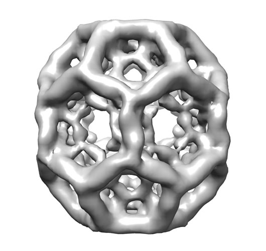 3442_imageClathrinD6coat.jpg
 http://ftp.pdbj.org/pub/emdb/structures/EMD-3442


 Sample components
 Processing
 Electron microscopy
