 Movie
Movie Controller

[English] 日本語
 Yorodumi
- EMDB-3291: Sub-tomogram averaging of Lassa virus glycoprotein spike from vir... -
+ Open data

- Basic information
| Entry | Database: EMDB / ID: EMD-3291 | |||||||||
|---|---|---|---|---|---|---|---|---|---|---|
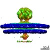
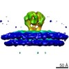
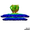
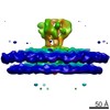
| Title | Sub-tomogram averaging of Lassa virus glycoprotein spike from virus-like particles at pH 7 | |||||||||
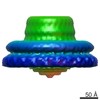
 Map data Map data | Sub-tomogram average of the glycoprotein spike trimer | |||||||||
 Sample Sample |
| |||||||||
 Keywords Keywords |  lassa virus / membrane protein / glycoprotein / receptor binding / membrane fusion lassa virus / membrane protein / glycoprotein / receptor binding / membrane fusion | |||||||||
| Biological species |  Lassa virus Lassa virus | |||||||||
| Method | subtomogram averaging / cryo EM / Resolution: 13.9 Å | |||||||||
 Authors Authors | Li S / Zhaoyang S / Pryce R / Parsy M-L / Fehling SK / Schlie K / Siebert CA / Garten W / Bowden TA / Strecker T / Huiskonen JT | |||||||||
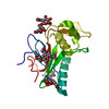
 Citation Citation | Journal: PLoS Pathog / Year: 2016 Title: Acidic pH-Induced Conformations and LAMP1 Binding of the Lassa Virus Glycoprotein Spike. Authors: Sai Li / Zhaoyang Sun / Rhys Pryce / Marie-Laure Parsy / Sarah K Fehling / Katrin Schlie / C Alistair Siebert / Wolfgang Garten / Thomas A Bowden / Thomas Strecker / Juha T Huiskonen /   Abstract: Lassa virus is an enveloped, bi-segmented RNA virus and the most prevalent and fatal of all Old World arenaviruses. Virus entry into the host cell is mediated by a tripartite surface spike complex, ...Lassa virus is an enveloped, bi-segmented RNA virus and the most prevalent and fatal of all Old World arenaviruses. Virus entry into the host cell is mediated by a tripartite surface spike complex, which is composed of two viral glycoprotein subunits, GP1 and GP2, and the stable signal peptide. Of these, GP1 binds to cellular receptors and GP2 catalyzes fusion between the viral envelope and the host cell membrane during endocytosis. The molecular structure of the spike and conformational rearrangements induced by low pH, prior to fusion, remain poorly understood. Here, we analyzed the three-dimensional ultrastructure of Lassa virus using electron cryotomography. Sub-tomogram averaging yielded a structure of the glycoprotein spike at 14-Å resolution. The spikes are trimeric, cover the virion envelope, and connect to the underlying matrix. Structural changes to the spike, following acidification, support a viral entry mechanism dependent on binding to the lysosome-resident receptor LAMP1 and further dissociation of the membrane-distal GP1 subunits. | |||||||||
| History |
|
- Structure visualization
| Movie |
 Movie viewer Movie viewer |
|---|---|
| Structure viewer | EM map: SurfViewMolmilJmol/JSmol |
| Supplemental images |

- Downloads & links
-EMDB archive
| Map data | emd_3291.map.gz | 7.5 MB | EMDB map data format | |
|---|---|---|---|---|
| Header (meta data) | emd-3291-v30.xmlemd-3291.xml | 10.9 KB 10.9 KB | Display Display | EMDB header |
| FSC (resolution estimation) | emd_3291_fsc.xml | 4.8 KB | Display | FSC data file |
| Images | emd_3291.tif | 1.2 MB | ||
| Archive directory |  http://ftp.pdbj.org/pub/emdb/structures/EMD-3291ftp://ftp.pdbj.org/pub/emdb/structures/EMD-3291 http://ftp.pdbj.org/pub/emdb/structures/EMD-3291ftp://ftp.pdbj.org/pub/emdb/structures/EMD-3291 | HTTPS FTP |
-Related structure data


-Links
| EMDB pages | EMDB (EBI/PDBe) / EMDataResource |
|---|
-Map
| File | Download / File: emd_3291.map.gz / Format: CCP4 / Size: 7.8 MB / Type: IMAGE STORED AS FLOATING POINT NUMBER (4 BYTES) | ||||||||||||||||||||||||||||||||||||||||||||||||||||||||||||
|---|---|---|---|---|---|---|---|---|---|---|---|---|---|---|---|---|---|---|---|---|---|---|---|---|---|---|---|---|---|---|---|---|---|---|---|---|---|---|---|---|---|---|---|---|---|---|---|---|---|---|---|---|---|---|---|---|---|---|---|---|---|
| Annotation | Sub-tomogram average of the glycoprotein spike trimer | ||||||||||||||||||||||||||||||||||||||||||||||||||||||||||||
| Voxel size | X=Y=Z: 2.7 Å | ||||||||||||||||||||||||||||||||||||||||||||||||||||||||||||
| Density |
| ||||||||||||||||||||||||||||||||||||||||||||||||||||||||||||
| Symmetry | Space group: 1 | ||||||||||||||||||||||||||||||||||||||||||||||||||||||||||||
| Details | EMDB XML:
CCP4 map header:
| ||||||||||||||||||||||||||||||||||||||||||||||||||||||||||||
-Supplemental data
- Sample components
-Entire : Purified Lassa virus VLPs at pH 7
| Entire | Name: Purified Lassa virus VLPs at pH 7 |
|---|---|
| Components |
|
-Supramolecule #1000: Purified Lassa virus VLPs at pH 7
| Supramolecule | Name: Purified Lassa virus VLPs at pH 7 / type: sample / ID: 1000 / Details: Unfixed virus-like particles / Number unique components: 1 |
|---|
-Supramolecule #1: Lassa virus
| Supramolecule | Name: Lassa virus / type: virus / ID: 1 / Name.synonym: Lassa mammarenavirus / NCBI-ID: 11620 / Sci species name: Lassa virus / Sci species strain: Josiah / Virus type: VIRUS-LIKE PARTICLE / Virus isolate: STRAIN / Virus enveloped: Yes / Virus empty: No / Syn species name: Lassa mammarenavirus |
|---|---|
| Host (natural) | Organism:  Mastomys (multimammate rats) / synonym: VERTEBRATES Mastomys (multimammate rats) / synonym: VERTEBRATES |
| Host system | Organism: Canis lupus (gray wolf) / Recombinant cell: Madin-Darby canine kidney |
-Experimental details
-Structure determination
| Method | cryo EM |
|---|---|
 Processing Processing | subtomogram averaging |
| Aggregation state | particle |
-Sample preparation
| Buffer | pH: 7.3 Details: 50 mM buffer of succinic acid, dihydrogen phosphate and glycine (2:7:7) |
|---|---|
| Grid | Details: Grids (Cflat CF-2/1-2C-T) were glow-discharged for 15 s. 6-nm gold particles were added. |
| Vitrification | Cryogen name: ETHANE-PROPANE MIXTURE / Chamber humidity: 80 % / Chamber temperature: 120 K / Instrument: GATAN CRYOPLUNGE 3 / Method: Blot for 3 seconds before plunging. |
- Electron microscopy
| Microscope | FEI POLARA 300 |
|---|---|
| Electron beam | Acceleration voltage: 300 kV / Electron source: FIELD EMISSION GUN |
| Electron optics | Calibrated magnification: 37037 / Illumination mode: FLOOD BEAM / Imaging mode: BRIGHT FIELDBright-field microscopy / Cs: 2.0 mm / Nominal defocus max: 3.7 µm / Nominal defocus min: 1.7 µm / Nominal magnification: 160000 |
| Specialist optics | Energy filter - Name: GIF QUANTUM LS / Energy filter - Lower energy threshold: 0.0 eV / Energy filter - Upper energy threshold: 20.0 eV |
| Sample stage | Specimen holder: Liquid nitrogen cooled / Specimen holder model: OTHER / Tilt series - Axis1 - Min angle: -45 ° / Tilt series - Axis1 - Max angle: 45 ° |
| Temperature | Min: 80 K / Max: 120 K |
| Alignment procedure | Legacy - Astigmatism: Objective lens astigmatism was corrected at 160,000 times magnification. |
| Details | Super-resolution counting mode |
| Date | May 15, 2014 |
| Image recording | Category: CCD / Film or detector model: GATAN K2 SUMMIT (4k x 4k) / Digitization - Sampling interval: 5 µm / Number real images: 31 / Average electron dose: 60 e/Å2 Details: Each image is a tilt series of 19 movies, acquired at 5 degree intervals. Each movie consists of 8 frames. |
| Experimental equipment |  Model: Tecnai Polara / Image courtesy: FEI Company |
-Image processing
| CTF correction | Details: Each tilted image |
|---|---|
| Final 3D classification | Number classes: 1 |
| Final reconstruction | Applied symmetry - Point group: C3 (3 fold cyclic) / Algorithm: OTHER / Resolution.type: BY AUTHOR / Resolution: 13.9 Å / Resolution method: OTHER / Software - Name: IMOD, Dynamo / Number subtomograms used: 2764 |
| Details | Subtomograms were picked manually. No C3 symmetry was imposed in the initial stages of refinement. |
| FSC plot (resolution estimation) |  |
