Journal: Cell Rep / Year: 2016 Title: Heterogeneous MAC Initiator and Pore Structures in a Lipid Bilayer by Phase-Plate Cryo-electron Tomography. Authors: Thomas H Sharp / Abraham J Koster / Piet Gros / Abstract: Pore formation in membranes is important for mammalian immune defense against invading bacteria. Induced by complement activation, the membrane attack complex (MAC) forms through sequential binding ...Pore formation in membranes is important for mammalian immune defense against invading bacteria. Induced by complement activation, the membrane attack complex (MAC) forms through sequential binding and membrane insertion of C5b6, C7, C8, and C9. Using cryo-electron tomography with a Volta phase plate and subtomogram averaging, we imaged C5b-7, C5b-8, and C5b-9 complexes and determined the C5b-9 pore structure in lipid bilayers. The in situ C5b-9 pore structure at 2.3-nm resolution reveals a 10- to 11.5-nm cone-shaped pore starting with C5b678 and multiple copies of C9 that is poorly closed, yielding a seam between C9 and C6 substituting for the shorter β strands in C6 and C7. However, large variations of composite pore complexes are apparent in subtomograms. Oligomerized initiator complexes C5b-7 and C5b-8 show stages of membrane binding, deformation, and perforation that yield ∼3.5-nm-wide pores. These data indicate a dynamic process of pore formation that likely adapts to biological membranes under attack.
History
Deposition
Dec 27, 2015
-
Header (metadata) release
Jan 27, 2016
-
Map release
Mar 30, 2016
-
Update
Mar 30, 2016
-
Current status
Mar 30, 2016
Processing site: PDBe / Status: Released
-
Structure visualization
Movie
Surface view with section colored by density value
Download / File: emd_3289.map.gz / Format: CCP4 / Size: 51.5 MB / Type: IMAGE STORED AS FLOATING POINT NUMBER (4 BYTES)
Annotation
Subtomogram average of the complement membrane attack complex
Voxel size
X=Y=Z: 2.861 Å
Density
Contour Level
By AUTHOR: 0.11 / Movie #1: 0.11
Minimum - Maximum
-0.1858658 - 0.41718206
Average (Standard dev.)
0.00101197 (±0.0297654)
Symmetry
Space group: 1
Details
EMDB XML:
Map geometry
Axis order
X
Y
Z
Origin
-120
-120
-120
Dimensions
240
240
240
Spacing
240
240
240
Cell
A=B=C: 686.64 Å α=β=γ: 90.0 °
CCP4 map header:
mode
Image stored as Reals
Å/pix. X/Y/Z
2.861
2.861
2.861
M x/y/z
240
240
240
origin x/y/z
0.000
0.000
0.000
length x/y/z
686.640
686.640
686.640
α/β/γ
90.000
90.000
90.000
MAP C/R/S
1
2
3
start NC/NR/NS
-120
-120
-120
NC/NR/NS
240
240
240
D min/max/mean
-0.186
0.417
0.001
-
Supplemental data
-
Sample components
-
Entire : Subtomogram average of the membrane attack complex pore in a lipi...
Entire
Name: Subtomogram average of the membrane attack complex pore in a lipid bilayer
Components
Sample: Subtomogram average of the membrane attack complex pore in a lipid bilayer
Protein or peptide: Complement component C5b6
Protein or peptide: Complement component C7
Protein or peptide: Complement component C8-alpha chain
Protein or peptide: Complement component C8-beta chain
Protein or peptide: Complement component C8-gamma chain
Protein or peptide: Complement component C9 chain
-
Supramolecule #1000: Subtomogram average of the membrane attack complex pore in a lipi...
Supramolecule
Name: Subtomogram average of the membrane attack complex pore in a lipid bilayer type: sample / ID: 1000 Oligomeric state: One copy of C5b, C6 and C7; one C8 heterotrimer; multiple copies of C9 Number unique components: 6
-
Macromolecule #1: Complement component C5b6
Macromolecule
Name: Complement component C5b6 / type: protein_or_peptide / ID: 1 / Name.synonym: C5b6 / Number of copies: 1 / Oligomeric state: Heterodimer / Recombinant expression: No
Source (natural)
Organism: Homo sapiens (human) / synonym: Human / Tissue: Serum
Molecular weight
Theoretical: 285 KDa
-
Macromolecule #2: Complement component C7
Macromolecule
Name: Complement component C7 / type: protein_or_peptide / ID: 2 / Name.synonym: C7 / Number of copies: 1 / Oligomeric state: Monomer / Recombinant expression: No
Source (natural)
Organism: Homo sapiens (human) / synonym: Human / Tissue: Serum
Name: Complement component C8-gamma chain / type: protein_or_peptide / ID: 5 / Name.synonym: C8gamma / Number of copies: 1 / Oligomeric state: Monomer / Recombinant expression: No
Source (natural)
Organism: Homo sapiens (human) / synonym: Human / Tissue: Serum
Molecular weight
Theoretical: 22 KDa
Sequence
UniProtKB: Complement component C8 gamma chain
-
Macromolecule #6: Complement component C9 chain
Macromolecule
Name: Complement component C9 chain / type: protein_or_peptide / ID: 6 / Name.synonym: C9 / Number of copies: 1 / Oligomeric state: Monomer / Recombinant expression: No
Source (natural)
Organism: Homo sapiens (human) / synonym: Human / Tissue: Serum
Molecular weight
Theoretical: 71 KDa
Sequence
UniProtKB: Complement component C9
-
Experimental details
-
Structure determination
Method
cryo EM
Processing
subtomogram averaging
Aggregation state
particle
-
Sample preparation
Concentration
0.241 mg/mL
Buffer
pH: 7.9 / Details: 10 mM Tris-HCl, 30 mM NaCl
Grid
Details: 300 mesh copper grids with lacey-carbon support, glow discharged.
Vitrification
Cryogen name: ETHANE / Chamber humidity: 95 % / Chamber temperature: 90.15 K / Instrument: LEICA EM GP Details: 6 ul sample pipetted onto freshly plasma-cleaned 300 mesh copper grids with lacey-carbon support Method: Blot for 6 seconds before plunging
-
Electron microscopy
Microscope
FEI TITAN KRIOS
Electron beam
Acceleration voltage: 300 kV / Electron source: FIELD EMISSION GUN
Specimen holder model: FEI TITAN KRIOS AUTOGRID HOLDER / Tilt series - Axis1 - Min angle: 60 ° / Tilt series - Axis1 - Max angle: 60 °
Details
Low-dose protocol used. Volta phase plate used
Date
Mar 10, 2015
Image recording
Category: CCD / Film or detector model: FEI FALCON II (4k x 4k) / Average electron dose: 1 e/Å2 / Details: Volta phase plate used
Experimental equipment
Model: Titan Krios / Image courtesy: FEI Company
-
Image processing
Final 3D classification
Number classes: 1
Final reconstruction
Applied symmetry - Point group: C1 (asymmetric) / Algorithm: OTHER / Resolution.type: BY AUTHOR / Resolution: 23.0 Å / Resolution method: OTHER / Software - Name: EMAN2 Details: Initial model was phase-randomized beyond 6 nm. Refinement was against a rotationally-averaged map. Number subtomograms used: 986
Details
Subtomograms were picked by hand
FSC plot (resolution estimation)
+
About Yorodumi
-
News
-
Feb 9, 2022. New format data for meta-information of EMDB entries
New format data for meta-information of EMDB entries
Version 3 of the EMDB header file is now the official format.
The previous official version 1.9 will be removed from the archive.
In the structure databanks used in Yorodumi, some data are registered as the other names, "COVID-19 virus" and "2019-nCoV". Here are the details of the virus and the list of structure data.
Jan 31, 2019. EMDB accession codes are about to change! (news from PDBe EMDB page)
EMDB accession codes are about to change! (news from PDBe EMDB page)
The allocation of 4 digits for EMDB accession codes will soon come to an end. Whilst these codes will remain in use, new EMDB accession codes will include an additional digit and will expand incrementally as the available range of codes is exhausted. The current 4-digit format prefixed with “EMD-” (i.e. EMD-XXXX) will advance to a 5-digit format (i.e. EMD-XXXXX), and so on. It is currently estimated that the 4-digit codes will be depleted around Spring 2019, at which point the 5-digit format will come into force.
The EM Navigator/Yorodumi systems omit the EMD- prefix.
Related info.:Q: What is EMD? / ID/Accession-code notation in Yorodumi/EM Navigator
Yorodumi is a browser for structure data from EMDB, PDB, SASBDB, etc.
This page is also the successor to EM Navigator detail page, and also detail information page/front-end page for Omokage search.
The word "yorodu" (or yorozu) is an old Japanese word meaning "ten thousand". "mi" (miru) is to see.
Related info.:EMDB / PDB / SASBDB / Comparison of 3 databanks / Yorodumi Search / Aug 31, 2016. New EM Navigator & Yorodumi / Yorodumi Papers / Jmol/JSmol / Function and homology information / Changes in new EM Navigator and Yorodumi
 Movie
Movie Controller


 Open data
 Basic information
 Map data
 Sample
 Keywords
 membrane attack complex / complement /
  Function and homology information
 Authors
 Citation
 Structure visualization
 Downloads & links
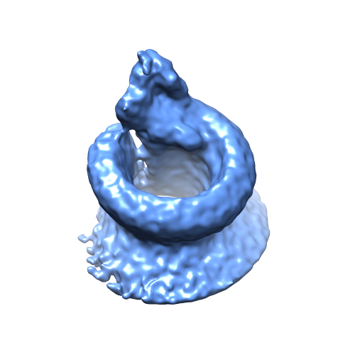 emd_3289.png
 http://ftp.pdbj.org/pub/emdb/structures/EMD-3289


 Sample components
 Processing
 Electron microscopy

