 Movie
Movie Controller

[English] 日本語
 Yorodumi
- EMDB-3131: Visualizing the Adsorption of Cyanophage P-SSP7 to the Marine Cya... -
+ Open data

- Basic information
| Entry | Database: EMDB / ID: EMD-3131 | |||||||||
|---|---|---|---|---|---|---|---|---|---|---|
| Title | Visualizing the Adsorption of Cyanophage P-SSP7 to the Marine Cyanobacterium Prochlorococcus MED4 by Electron Cryo-Tomography | |||||||||
 Map data Map data | pre-infection conformation of P-SSP7 bacteriophage | |||||||||
 Sample Sample |
| |||||||||
 Keywords Keywords |  cryo-electron tomography / bacteriophage / cyanobacteria cryo-electron tomography / bacteriophage / cyanobacteria | |||||||||
| Biological species |  Prochlorococcus phage P-SSP7 (virus) Prochlorococcus phage P-SSP7 (virus) | |||||||||
| Method | subtomogram averaging / cryo EM | |||||||||
 Authors Authors | Murata K / Zhang Q / Fu C / Liu X / Sullivan M / Coleman M / Osburne M / Schmid MF / Chisholm S / Chiu W | |||||||||
 Citation Citation | Journal: Sci Rep / Year: 2017 Title: Visualizing Adsorption of Cyanophage P-SSP7 onto Marine Prochlorococcus. Authors: Kazuyoshi Murata / Qinfen Zhang / Jesús Gerardo Galaz-Montoya / Caroline Fu / Maureen L Coleman / Marcia S Osburne / Michael F Schmid / Matthew B Sullivan / Sallie W Chisholm / Wah Chiu /    Abstract: Marine cyanobacteria perform roughly a quarter of global carbon fixation, and cyanophages that infect them liberate some of this carbon during infection and cell lysis. Studies of the cyanobacterium ...Marine cyanobacteria perform roughly a quarter of global carbon fixation, and cyanophages that infect them liberate some of this carbon during infection and cell lysis. Studies of the cyanobacterium Prochlorococcus MED4 and its associated cyanophage P-SSP7 have revealed complex gene expression dynamics once infection has begun, but the initial cyanophage-host interactions remain poorly understood. Here, we used single particle cryo-electron tomography (cryo-ET) to investigate cyanophage-host interactions in this model system, based on 170 cyanophage-to-host adsorption events. Subtomogram classification and averaging revealed three main conformations characterized by different angles between the phage tail and the cell surface. Namely, phage tails were (i) parallel to, (ii) ~45 degrees to, or (iii) perpendicular to the cell surface. Furthermore, different conformations of phage tail fibers correlated with the aforementioned orientations of the tails. We also observed density beyond the tail tip in vertically-oriented phages that had penetrated the cell wall, capturing the final stage of adsorption. Together, our data provide a quantitative characterization of the orientation of phages as they adsorb onto cells, and suggest that cyanophages that abut their cellular targets are only transiently in the "perpendicular" orientation required for successful infection. | |||||||||
| History |
|
- Structure visualization
| Movie |
 Movie viewer Movie viewer |
|---|---|
| Structure viewer | EM map: SurfViewMolmilJmol/JSmol |
| Supplemental images |

- Downloads & links
-EMDB archive
| Map data | emd_3131.map.gz | 9.4 MB | EMDB map data format | |
|---|---|---|---|---|
| Header (meta data) | emd-3131-v30.xmlemd-3131.xml | 8.8 KB 8.8 KB | Display Display | EMDB header |
| Images | 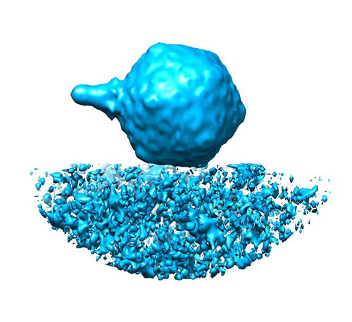 EMD-3131.jpg EMD-3131.jpg | 178.9 KB | ||
| Archive directory |  http://ftp.pdbj.org/pub/emdb/structures/EMD-3131ftp://ftp.pdbj.org/pub/emdb/structures/EMD-3131 http://ftp.pdbj.org/pub/emdb/structures/EMD-3131ftp://ftp.pdbj.org/pub/emdb/structures/EMD-3131 | HTTPS FTP |
-Related structure data


-Links
| EMDB pages | EMDB (EBI/PDBe) / EMDataResource |
|---|
-Map
| File | Download / File: emd_3131.map.gz / Format: CCP4 / Size: 21.7 MB / Type: IMAGE STORED AS FLOATING POINT NUMBER (4 BYTES) | ||||||||||||||||||||||||||||||||||||||||||||||||||||||||||||
|---|---|---|---|---|---|---|---|---|---|---|---|---|---|---|---|---|---|---|---|---|---|---|---|---|---|---|---|---|---|---|---|---|---|---|---|---|---|---|---|---|---|---|---|---|---|---|---|---|---|---|---|---|---|---|---|---|---|---|---|---|---|
| Annotation | pre-infection conformation of P-SSP7 bacteriophage | ||||||||||||||||||||||||||||||||||||||||||||||||||||||||||||
| Voxel size | X=Y=Z: 9.36 Å | ||||||||||||||||||||||||||||||||||||||||||||||||||||||||||||
| Density |
| ||||||||||||||||||||||||||||||||||||||||||||||||||||||||||||
| Symmetry | Space group: 1 | ||||||||||||||||||||||||||||||||||||||||||||||||||||||||||||
| Details | EMDB XML:
CCP4 map header:
| ||||||||||||||||||||||||||||||||||||||||||||||||||||||||||||
-Supplemental data
- Sample components
-Entire : Pre-infection comformation of Prochlorococcus phage P-SSP7
| Entire | Name: Pre-infection comformation of Prochlorococcus phage P-SSP7 |
|---|---|
| Components |
|
-Supramolecule #1000: Pre-infection comformation of Prochlorococcus phage P-SSP7
| Supramolecule | Name: Pre-infection comformation of Prochlorococcus phage P-SSP7 type: sample / ID: 1000 / Number unique components: 1 |
|---|
-Supramolecule #1: Prochlorococcus phage P-SSP7
| Supramolecule | Name: Prochlorococcus phage P-SSP7 / type: virus / ID: 1 / NCBI-ID: 268748 / Sci species name: Prochlorococcus phage P-SSP7 / Sci species strain: P-SSP7 / Virus type: VIRION / Virus isolate: STRAIN / Virus enveloped: No / Virus empty: No |
|---|---|
| Host (natural) | Organism:  Prochlorococcus (bacteria) / Strain: MED4 / synonym: BACTERIA(EUBACTERIA) Prochlorococcus (bacteria) / Strain: MED4 / synonym: BACTERIA(EUBACTERIA) |
| Virus shell | Shell ID: 1 / Diameter: 655 Å / T number (triangulation number): 7 |
-Experimental details
-Structure determination
| Method | cryo EM |
|---|---|
 Processing Processing | subtomogram averaging |
| Aggregation state | cell |
-Sample preparation
| Buffer | pH: 7.5 Details: 100 mM Tris-HCl (pH 7.5), 100 mM MgSO4, and 30 mM NaCl |
|---|---|
| Grid | Details: R3.5/1 Quantifoil |
| Vitrification | Cryogen name: ETHANE / Chamber humidity: 40 % / Chamber temperature: 120 K / Instrument: LEICA EM CPC Method: Grids with sample solution were quickly frozen in liquid ethane. |
- Electron microscopy
| Microscope | JEOL 3200FSC |
|---|---|
| Electron beam | Acceleration voltage: 300 kV / Electron source: FIELD EMISSION GUN |
| Electron optics | Illumination mode: FLOOD BEAM / Imaging mode: BRIGHT FIELDBright-field microscopy / Cs: 4.1 mm / Nominal defocus max: 5.0 µm / Nominal defocus min: 3.0 µm / Nominal magnification: 20000 |
| Specialist optics | Energy filter - Name: Omega / Energy filter - Lower energy threshold: 0.0 eV / Energy filter - Upper energy threshold: 30.0 eV |
| Sample stage | Specimen holder model: JEOL 3200FSC CRYOHOLDER / Tilt series - Axis1 - Min angle: 0 ° / Tilt series - Axis1 - Max angle: 62 ° |
| Temperature | Average: 90 K |
| Date | May 16, 2008 |
| Image recording | Category: CCD / Film or detector model: GATAN ULTRASCAN 4000 (4k x 4k) / Average electron dose: 80 e/Å2 / Bits/pixel: 16 |
-Image processing
| Final reconstruction | Applied symmetry - Point group: C1 (asymmetric) / Algorithm: OTHER / Resolution method: OTHER / Software - Name: EMAN1 / Number subtomograms used: 44 |
|---|
