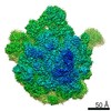
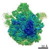
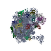
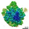
Journal: Structure / Year: 2016 Title: The Structure of the Mouse Serotonin 5-HT3 Receptor in Lipid Vesicles. Authors: Mikhail Kudryashev / Daniel Castaño-Díez / Cédric Deluz / Gherici Hassaine / Luigino Grasso / Alexandra Graf-Meyer / Horst Vogel / Henning Stahlberg / Abstract: The function of membrane proteins is best understood if their structure in the lipid membrane is known. Here, we determined the structure of the mouse serotonin 5-HT3 receptor inserted in lipid ...The function of membrane proteins is best understood if their structure in the lipid membrane is known. Here, we determined the structure of the mouse serotonin 5-HT3 receptor inserted in lipid bilayers to a resolution of 12 Å without stabilizing antibodies by cryo electron tomography and subtomogram averaging. The reconstruction reveals protein secondary structure elements in the transmembrane region, the extracellular pore, and the transmembrane channel pathway, showing an overall similarity to the available X-ray model of the truncated 5-HT3 receptor determined in the presence of a stabilizing nanobody. Structural analysis of the 5-HT3 receptor embedded in a lipid bilayer allowed the position of the membrane to be determined. Interactions between the densely packed receptors in lipids were visualized, revealing that the interactions were maintained by the short horizontal helices. In combination with methodological improvements, our approach enables the structural analysis of membrane proteins in response to voltage and ligand gating.
History
Deposition
Jul 24, 2015
-
Header (metadata) release
Aug 19, 2015
-
Map release
Jan 13, 2016
-
Update
Jan 27, 2016
-
Current status
Jan 27, 2016
Processing site: PDBe / Status: Released
-
Structure visualization
Movie
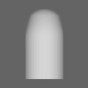
Surface view with section colored by density value
EMPIAR-10046 (Title: Cryo electron tomography of mouse 5-HT3 receptors in lipid vesicles Data size: 158.8 Data #1: Tilt series of the 5-HT3 receptors reconstituted to lipid vesicles [class averages])
Cryogen name: ETHANE / Chamber humidity: 90 % / Chamber temperature: 90 K / Instrument: LEICA EM GP / Method: Blot for 2 second before plunding
-
Electron microscopy
Microscope
FEI TITAN KRIOS
Alignment procedure
Legacy - Astigmatism: corrected at high magnification
Details
tomograms acquired without the energy filtration
Date
Dec 1, 2013
Image recording
Category: CCD / Film or detector model: GATAN K2 (4k x 4k) / Number real images: 46 / Average electron dose: 41 e/Å2 / Details: 46 tomograms were selected for processing
Electron beam
Acceleration voltage: 300 kV / Electron source: FIELD EMISSION GUN
Specimen holder model: FEI TITAN KRIOS AUTOGRID HOLDER / Tilt series - Axis1 - Min angle: -60 ° / Tilt series - Axis1 - Max angle: 60 °
Experimental equipment
Model: Titan Krios / Image courtesy: FEI Company
-
Image processing
Details
Particles were automatically picked from the surface of the vesicles in CTF-corrected cryo-electron tomograms. After a rough alignment deep classification into 20 classes was performed including 4 neighbouring pentamers in the alignment mask. Finally the central pentamers from each class were cropped, aligned and averaged resulting in a final structure.
Final reconstruction
Applied symmetry - Point group: C5 (5 fold cyclic) / Resolution.type: BY AUTHOR / Resolution: 12.0 Å / Resolution method: OTHER / Software - Name: IMOD, Dynamo / Number subtomograms used: 16000
CTF correction
Details: ctfplotter
Final 3D classification
Number classes: 20
+
About Yorodumi
-
News
-
Feb 9, 2022. New format data for meta-information of EMDB entries
New format data for meta-information of EMDB entries
Version 3 of the EMDB header file is now the official format.
The previous official version 1.9 will be removed from the archive.
In the structure databanks used in Yorodumi, some data are registered as the other names, "COVID-19 virus" and "2019-nCoV". Here are the details of the virus and the list of structure data.
Jan 31, 2019. EMDB accession codes are about to change! (news from PDBe EMDB page)
EMDB accession codes are about to change! (news from PDBe EMDB page)
The allocation of 4 digits for EMDB accession codes will soon come to an end. Whilst these codes will remain in use, new EMDB accession codes will include an additional digit and will expand incrementally as the available range of codes is exhausted. The current 4-digit format prefixed with “EMD-” (i.e. EMD-XXXX) will advance to a 5-digit format (i.e. EMD-XXXXX), and so on. It is currently estimated that the 4-digit codes will be depleted around Spring 2019, at which point the 5-digit format will come into force.
The EM Navigator/Yorodumi systems omit the EMD- prefix.
Related info.:Q: What is EMD? / ID/Accession-code notation in Yorodumi/EM Navigator
Yorodumi is a browser for structure data from EMDB, PDB, SASBDB, etc.
This page is also the successor to EM Navigator detail page, and also detail information page/front-end page for Omokage search.
The word "yorodu" (or yorozu) is an old Japanese word meaning "ten thousand". "mi" (miru) is to see.
Related info.:EMDB / PDB / SASBDB / Comparison of 3 databanks / Yorodumi Search / Aug 31, 2016. New EM Navigator & Yorodumi / Yorodumi Papers / Jmol/JSmol / Function and homology information / Changes in new EM Navigator and Yorodumi
 Movie
Movie Controller

 Yorodumi
 Open data
 Basic information
 Map data
 Sample
 Function and homology information
 Authors
 Citation

 Structure visualization
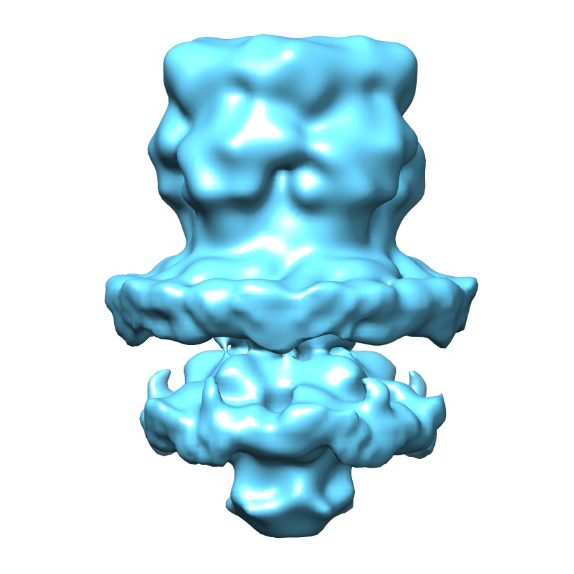
 Downloads & links
 http://ftp.pdbj.org/pub/emdb/structures/EMD-3108


 Z
Z Y
Y X
X


 Sample components
 Homo sapiens (human) / Recombinant cell: T-REx-293 cells / Recombinant plasmid: pcDNA5/TO
 Processing
 Electron microscopy
 FIELD EMISSION GUN
