 Movie
Movie Controller


+ Open data

- Basic information
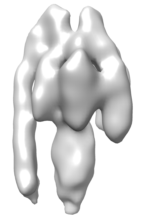
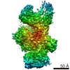
| Entry | Database: EMDB / ID: EMD-2982 | |||||||||
|---|---|---|---|---|---|---|---|---|---|---|
| Title | Sub-tomogram average of a mammalian F-type ATP synthase monomer | |||||||||
 Map data Map data | Sub-tomogram average of bovine F-type ATP synthase in a 2D crystal | |||||||||
 Sample Sample |
| |||||||||
 Keywords Keywords | F-type ATP synthase / Mitochondria Bovine heart | |||||||||
| Biological species |   Bos taurus (cattle) Bos taurus (cattle) | |||||||||
| Method | subtomogram averaging / cryo EM / negative staining / Resolution: 24.0 Å | |||||||||
 Authors Authors | Jiko C / Davies KM / Shinzawa-Itoh K / Tani K / Maeda S / Mills DJ / Tsukihara T / Fujiyoshi Y / Kuehlbrandt W / Gerle C | |||||||||
 Citation Citation | Journal: Elife / Year: 2015 Title: Bovine F1Fo ATP synthase monomers bend the lipid bilayer in 2D membrane crystals. Authors: Chimari Jiko / Karen M Davies / Kyoko Shinzawa-Itoh / Kazutoshi Tani / Shintaro Maeda / Deryck J Mills / Tomitake Tsukihara / Yoshinori Fujiyoshi / Werner Kühlbrandt / Christoph Gerle /   Abstract: We have used a combination of electron cryo-tomography, subtomogram averaging, and electron crystallographic image processing to analyse the structure of intact bovine F(1)F(o) ATP synthase in 2D ...We have used a combination of electron cryo-tomography, subtomogram averaging, and electron crystallographic image processing to analyse the structure of intact bovine F(1)F(o) ATP synthase in 2D membrane crystals. ATPase assays and mass spectrometry analysis of the 2D crystals confirmed that the enzyme complex was complete and active. The structure of the matrix-exposed region was determined at 24 Å resolution by subtomogram averaging and repositioned into the tomographic volume to reveal the crystal packing. F(1)F(o) ATP synthase complexes are inclined by 16° relative to the crystal plane, resulting in a zigzag topology of the membrane and indicating that monomeric bovine heart F(1)F(o) ATP synthase by itself is sufficient to deform lipid bilayers. This local membrane curvature is likely to be instrumental in the formation of ATP synthase dimers and dimer rows, and thus for the shaping of mitochondrial cristae. | |||||||||
| History |
|
- Structure visualization
| Movie |
 Movie viewer Movie viewer |
|---|---|
| Structure viewer | EM map: SurfViewMolmilJmol/JSmol |
| Supplemental images |
- Downloads & links
-EMDB archive
| Map data | emd_2982.map.gz | 339 KB | EMDB map data format | |
|---|---|---|---|---|
| Header (meta data) | emd-2982-v30.xmlemd-2982.xml | 11.3 KB 11.3 KB | Display Display | EMDB header |
| Images |  EMD-2982.png EMD-2982.png | 90.5 KB | ||
| Archive directory |  http://ftp.pdbj.org/pub/emdb/structures/EMD-2982ftp://ftp.pdbj.org/pub/emdb/structures/EMD-2982 http://ftp.pdbj.org/pub/emdb/structures/EMD-2982ftp://ftp.pdbj.org/pub/emdb/structures/EMD-2982 | HTTPS FTP |
-Related structure data
| Similar structure data | |
|---|---|
| EM raw data | EMPIAR-10027 (Title: Sub-tomogram average of a mammalian F-type ATP synthase monomer Data size: 8.9 / Data #1: 2dxtal_A [class averages] / Data #2: 2dxtal_B [class averages]) |


-Links
| EMDB pages | EMDB (EBI/PDBe) / EMDataResource |
|---|
-Map
| File | Download / File: emd_2982.map.gz / Format: CCP4 / Size: 429.7 KB / Type: IMAGE STORED AS FLOATING POINT NUMBER (4 BYTES) | ||||||||||||||||||||||||||||||||||||||||||||||||||||||||||||
|---|---|---|---|---|---|---|---|---|---|---|---|---|---|---|---|---|---|---|---|---|---|---|---|---|---|---|---|---|---|---|---|---|---|---|---|---|---|---|---|---|---|---|---|---|---|---|---|---|---|---|---|---|---|---|---|---|---|---|---|---|---|
| Annotation | Sub-tomogram average of bovine F-type ATP synthase in a 2D crystal | ||||||||||||||||||||||||||||||||||||||||||||||||||||||||||||
| Voxel size | X=Y=Z: 3.3 Å | ||||||||||||||||||||||||||||||||||||||||||||||||||||||||||||
| Density |
| ||||||||||||||||||||||||||||||||||||||||||||||||||||||||||||
| Symmetry | Space group: 1 | ||||||||||||||||||||||||||||||||||||||||||||||||||||||||||||
| Details | EMDB XML:
CCP4 map header:
| ||||||||||||||||||||||||||||||||||||||||||||||||||||||||||||
-Supplemental data
- Sample components
-Entire : 2D crystal of bovine F-type ATP synthase monomers
| Entire | Name: 2D crystal of bovine F-type ATP synthase monomers |
|---|---|
| Components |
|
-Supramolecule #1000: 2D crystal of bovine F-type ATP synthase monomers
| Supramolecule | Name: 2D crystal of bovine F-type ATP synthase monomers / type: sample / ID: 1000 Details: 2D Membrane crystals lipids for reconstitution: 1,2-dimyristoyl-sn-glycero-3-phosphocholine Oligomeric state: 1 / Number unique components: 1 |
|---|---|
| Molecular weight | Theoretical: 600 KDa |
-Macromolecule #1: F-type ATP synthase
| Macromolecule | Name: F-type ATP synthase / type: protein_or_peptide / ID: 1 / Number of copies: 1 / Oligomeric state: monomer / Recombinant expression: No |
|---|---|
| Source (natural) | Organism: Bos taurus (cattle) / synonym: cattle / Tissue: Heart / Organelle: Mitochondria / Location in cell: Mitochondria |
| Molecular weight | Theoretical: 600 KDa |
-Experimental details
-Structure determination
| Method | negative staining, cryo EM |
|---|---|
 Processing Processing | subtomogram averaging |
| Aggregation state | 2D array |
-Sample preparation
| Concentration | 10 mg/mL |
|---|---|
| Buffer | pH: 8.2 Details: 40mM Tris-HCL pH8.2 100mM NaCl 0.02%(w/v)NaN3, 0.5mM ADP, 5mM MgCl2, 0.1mM DTT, 0.1mM EDTA |
| Staining | Type: NEGATIVE Details: Plunge frozen in liquid ethane using home-made freezing device |
| Grid | Details: Glow discharged R2/2, 300 copper mesh quantifoil grids |
| Vitrification | Cryogen name: ETHANE / Chamber temperature: 100 K / Instrument: HOMEMADE PLUNGER Method: 2D crystals were mixed 1:1 (v/v) with 6nm collodial gold particles. Three microliters of protein/gold sample was applied to R2/2 300 copper mesh quantifoil grids. Access liquid was removed by ...Method: 2D crystals were mixed 1:1 (v/v) with 6nm collodial gold particles. Three microliters of protein/gold sample was applied to R2/2 300 copper mesh quantifoil grids. Access liquid was removed by blotting for 3s with Whatman #4 paper before plunge-freezing in liquid ethane |
| Details | 10mg/ml ATP synthase were mixed with 1,2-dimyristoyl-sn-glycero-3-phosphocholine (Avanti Polar Lipids) using a lipid to protein ratio of 0.2. Detergent was removed by dialysis using 20 microliter Hampton dialysis buttons and 15kDa membrane cuttoff. |
- Electron microscopy
| Microscope | FEI TITAN KRIOS |
|---|---|
| Electron beam | Acceleration voltage: 300 kV / Electron source: FIELD EMISSION GUN |
| Electron optics | Calibrated magnification: 14942 / Illumination mode: FLOOD BEAM / Imaging mode: BRIGHT FIELDBright-field microscopy / Cs: 2.7 mm / Nominal defocus max: 3.0 µm / Nominal defocus min: 2.0 µm / Nominal magnification: 42000 |
| Specialist optics | Energy filter - Name: quantum / Energy filter - Lower energy threshold: 0.0 eV / Energy filter - Upper energy threshold: 20.0 eV |
| Sample stage | Specimen holder: Nitrogen cooled / Specimen holder model: FEI TITAN KRIOS AUTOGRID HOLDER / Tilt series - Axis1 - Min angle: -60 ° / Tilt series - Axis1 - Max angle: 60 ° |
| Temperature | Min: 80 K / Max: 100 K / Average: 90 K |
| Alignment procedure | Legacy - Astigmatism: Objection lend astigmation was corrected on K2 at magnification used for imaging. |
| Details | tomographic tilt series |
| Date | Feb 1, 2013 |
| Image recording | Category: CCD / Film or detector model: GATAN K2 (4k x 4k) / Number real images: 80 / Average electron dose: 60 e/Å2 |
| Experimental equipment |  Model: Titan Krios / Image courtesy: FEI Company |
-Image processing
| Crystal parameters | Unit cell - A: 179.1 Å / Unit cell - B: 171.4 Å / Unit cell - γ: 94.9 ° / Plane group: P 1 |
|---|---|
| CTF correction | Details: IMOD |
| Final reconstruction | Applied symmetry - Point group: C1 (asymmetric) / Resolution.type: BY AUTHOR / Resolution: 24.0 Å / Resolution method: OTHER / Software - Name: IMOD / Number subtomograms used: 2100 |
| Details | Subtomograms were selected manually and averaged using PEET (Particle Estimation for Electron Tomography, Boulder) |
