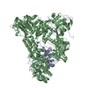
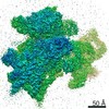
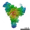
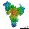
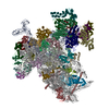
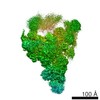
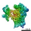
Journal: Nature / Year: 2015 Title: The architecture of the spliceosomal U4/U6.U5 tri-snRNP. Authors: Thi Hoang Duong Nguyen / Wojciech P Galej / Xiao-chen Bai / Christos G Savva / Andrew J Newman / Sjors H W Scheres / Kiyoshi Nagai / Abstract: U4/U6.U5 tri-snRNP is a 1.5-megadalton pre-assembled spliceosomal complex comprising U5 small nuclear RNA (snRNA), extensively base-paired U4/U6 snRNAs and more than 30 proteins, including the key ...U4/U6.U5 tri-snRNP is a 1.5-megadalton pre-assembled spliceosomal complex comprising U5 small nuclear RNA (snRNA), extensively base-paired U4/U6 snRNAs and more than 30 proteins, including the key components Prp8, Brr2 and Snu114. The tri-snRNP combines with a precursor messenger RNA substrate bound to U1 and U2 small nuclear ribonucleoprotein particles (snRNPs), and transforms into a catalytically active spliceosome after extensive compositional and conformational changes triggered by unwinding of the U4 and U6 (U4/U6) snRNAs. Here we use cryo-electron microscopy single-particle reconstruction of Saccharomyces cerevisiae tri-snRNP at 5.9 Å resolution to reveal the essentially complete organization of its RNA and protein components. The single-stranded region of U4 snRNA between its 3' stem-loop and the U4/U6 snRNA stem I is loaded into the Brr2 helicase active site ready for unwinding. Snu114 and the amino-terminal domain of Prp8 position U5 snRNA to insert its loop I, which aligns the exons for splicing, into the Prp8 active site cavity. The structure provides crucial insights into the activation process and the active site of the spliceosome.
History
Deposition
Mar 29, 2015
-
Header (metadata) release
May 6, 2015
-
Map release
Jul 1, 2015
-
Update
Jul 1, 2015
-
Current status
Jul 1, 2015
Processing site: PDBe / Status: Released
-
Structure visualization
Movie
Surface view with section colored by density value
Category: CCD / Film or detector model: FEI FALCON II (4k x 4k) / Number real images: 2035 / Average electron dose: 40 e/Å2 Details: Every image is the average of sixteen frames recorded by the direct electron detector
Experimental equipment
Model: Tecnai Polara / Image courtesy: FEI Company
+
Image processing
CTF correction
Details: each particle
Final reconstruction
Applied symmetry - Point group: C1 (asymmetric) / Resolution.type: BY AUTHOR / Resolution: 5.9 Å / Resolution method: OTHER / Software - Name: CTFFIND3, RELION Details: To correct for beam-induced movements, the 16 video frames for each micrograph were first aligned using whole-image motion correction. Second, particle based beam-induced movement correction ...Details: To correct for beam-induced movements, the 16 video frames for each micrograph were first aligned using whole-image motion correction. Second, particle based beam-induced movement correction was performed using statistical movie processing in RELION. Number images used: 179079
In the structure databanks used in Yorodumi, some data are registered as the other names, "COVID-19 virus" and "2019-nCoV". Here are the details of the virus and the list of structure data.
Jan 31, 2019. EMDB accession codes are about to change! (news from PDBe EMDB page)
EMDB accession codes are about to change! (news from PDBe EMDB page)
The allocation of 4 digits for EMDB accession codes will soon come to an end. Whilst these codes will remain in use, new EMDB accession codes will include an additional digit and will expand incrementally as the available range of codes is exhausted. The current 4-digit format prefixed with “EMD-” (i.e. EMD-XXXX) will advance to a 5-digit format (i.e. EMD-XXXXX), and so on. It is currently estimated that the 4-digit codes will be depleted around Spring 2019, at which point the 5-digit format will come into force.
The EM Navigator/Yorodumi systems omit the EMD- prefix.
Related info.:Q: What is EMD? / ID/Accession-code notation in Yorodumi/EM Navigator
Yorodumi is a browser for structure data from EMDB, PDB, SASBDB, etc.
This page is also the successor to EM Navigator detail page, and also detail information page/front-end page for Omokage search.
The word "yorodu" (or yorozu) is an old Japanese word meaning "ten thousand". "mi" (miru) is to see.
Related info.:EMDB / PDB / SASBDB / Comparison of 3 databanks / Yorodumi Search / Aug 31, 2016. New EM Navigator & Yorodumi / Yorodumi Papers / Jmol/JSmol / Function and homology information / Changes in new EM Navigator and Yorodumi
 Movie
Movie Controller


 Open data
 Basic information
 Map data
 Sample
 Keywords
 Spliceosome /
 Authors
 Citation
 Structure visualization
 Movie viewer
 Downloads & links
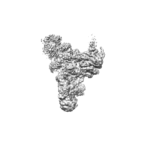 emd_2966.png
 http://ftp.pdbj.org/pub/emdb/structures/EMD-2966
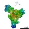

 Sample components
 Processing
 Electron microscopy