 Movie
Movie Controller


+ Open data

- Basic information
| Entry | Database: EMDB / ID: EMD-2865 | |||||||||
|---|---|---|---|---|---|---|---|---|---|---|
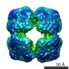
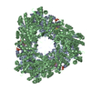
| Title | Architecture of the yeast pontin-reptin complex | |||||||||
 Map data Map data | 3D reconstruction of the yeast pontin-reptin complex | |||||||||
 Sample Sample |
| |||||||||
 Keywords Keywords | Rvb1 / Rvb2 / Tip48 / Tip49 / pontin /  reptin / Ino80 / AAA+ reptin / Ino80 / AAA+ | |||||||||
| Function / homology | TIP49, P-loop domain / Ino80 complex Function and homology information Function and homology information | |||||||||
| Biological species |  Saccharomyces cerevisiae (brewer's yeast) Saccharomyces cerevisiae (brewer's yeast) | |||||||||
| Method | single particle reconstruction / cryo EM / Resolution: 13.6 Å | |||||||||
 Authors Authors | Torreira E / Jha S / Lopez-Blanco JR / Arias-Palomo E / Chacon P / Canas C / Ayora S / Dutta A / Llorca O | |||||||||
 Citation Citation | Journal: Structure / Year: 2008 Title: Architecture of the pontin/reptin complex, essential in the assembly of several macromolecular complexes. Authors: Eva Torreira / Sudhakar Jha / José R López-Blanco / Ernesto Arias-Palomo / Pablo Chacón / Cristina Cañas / Sylvia Ayora / Anindya Dutta / Oscar Llorca /  Abstract: Pontin and reptin belong to the AAA+ family, and they are essential for the structural integrity and catalytic activity of several chromatin remodeling complexes. They are also indispensable for the ...Pontin and reptin belong to the AAA+ family, and they are essential for the structural integrity and catalytic activity of several chromatin remodeling complexes. They are also indispensable for the assembly of several ribonucleoprotein complexes, including telomerase. Here, we propose a structural model of the yeast pontin/reptin complex based on a cryo-electron microscopy reconstruction at 13 A. Pontin/reptin hetero-dodecamers were purified from in vivo assembled complexes forming a double ring. Two rings interact through flexible domains projecting from each hexamer, constituting an atypical asymmetric form of oligomerization. These flexible domains and the AAA+ cores reveal significant conformational changes when compared with the crystal structure of human pontin that generate enlarged channels. This structure of endogenously assembled pontin/reptin complexes is different than previously described structures, suggesting that pontin and reptin could acquire distinct structural states to regulate their broad functions as molecular motors and scaffolds for nucleic acids and proteins. | |||||||||
| History |
|
- Structure visualization
| Movie |
Movie viewer |
|---|---|
| Structure viewer | EM map: SurfViewMolmilJmol/JSmol |
| Supplemental images |

- Downloads & links
-EMDB archive
| Map data | emd_2865.map.gz | 711.8 KB | EMDB map data format | |
|---|---|---|---|---|
| Header (meta data) | emd-2865-v30.xmlemd-2865.xml | 11.7 KB 11.7 KB | Display Display | EMDB header |
| Images |  emd-2865.gif emd-2865.gif | 92.4 KB | ||
| Archive directory |  http://ftp.pdbj.org/pub/emdb/structures/EMD-2865ftp://ftp.pdbj.org/pub/emdb/structures/EMD-2865 http://ftp.pdbj.org/pub/emdb/structures/EMD-2865ftp://ftp.pdbj.org/pub/emdb/structures/EMD-2865 | HTTPS FTP |
-Related structure data
| Similar structure data |
|---|


-Links
| EMDB pages | EMDB (EBI/PDBe) / EMDataResource |
|---|
-Map
| File | Download / File: emd_2865.map.gz / Format: CCP4 / Size: 4.2 MB / Type: IMAGE STORED AS FLOATING POINT NUMBER (4 BYTES) | ||||||||||||||||||||||||||||||||||||||||||||||||||||||||||||||||||||
|---|---|---|---|---|---|---|---|---|---|---|---|---|---|---|---|---|---|---|---|---|---|---|---|---|---|---|---|---|---|---|---|---|---|---|---|---|---|---|---|---|---|---|---|---|---|---|---|---|---|---|---|---|---|---|---|---|---|---|---|---|---|---|---|---|---|---|---|---|---|
| Annotation | 3D reconstruction of the yeast pontin-reptin complex | ||||||||||||||||||||||||||||||||||||||||||||||||||||||||||||||||||||
| Voxel size | X=Y=Z: 2.12 Å | ||||||||||||||||||||||||||||||||||||||||||||||||||||||||||||||||||||
| Density |
| ||||||||||||||||||||||||||||||||||||||||||||||||||||||||||||||||||||
| Symmetry | Space group: 1 | ||||||||||||||||||||||||||||||||||||||||||||||||||||||||||||||||||||
| Details | EMDB XML:
CCP4 map header:
| ||||||||||||||||||||||||||||||||||||||||||||||||||||||||||||||||||||
-Supplemental data
- Sample components
-Entire : Complex between the proteins pontin and reptin
| Entire | Name: Complex between the proteins pontin and reptin |
|---|---|
| Components |
|
-Supramolecule #1000: Complex between the proteins pontin and reptin
| Supramolecule | Name: Complex between the proteins pontin and reptin / type: sample / ID: 1000 Oligomeric state: Dodecamer composed of 6 subunits of pontin and 6 subunits of reptin Number unique components: 2 |
|---|---|
| Molecular weight | Theoretical: 650 KDa |
-Macromolecule #1: RuvB-like 1
| Macromolecule | Name: RuvB-like 1 / type: protein_or_peptide / ID: 1 / Name.synonym: Pontin / Number of copies: 6 / Oligomeric state: Hexamer / Recombinant expression: Yes |
|---|---|
| Source (natural) | Organism: Saccharomyces cerevisiae (brewer's yeast) / synonym: Baker's yeast |
| Molecular weight | Theoretical: 540 KDa |
| Recombinant expression | Organism: Insect cells using recombinant baculovirus |
| Sequence | GO: Ino80 complex / InterPro: TIP49, P-loop domain |
-Macromolecule #2: RuvB-like 2
| Macromolecule | Name: RuvB-like 2 / type: protein_or_peptide / ID: 2 / Name.synonym: Reptin / Number of copies: 6 / Oligomeric state: Hexamer / Recombinant expression: Yes |
|---|---|
| Source (natural) | Organism: Saccharomyces cerevisiae (brewer's yeast) / synonym: Baker's yeast |
| Molecular weight | Theoretical: 540 KDa |
| Recombinant expression | Organism: Insect cells using recombinant baculovirus |
| Sequence | GO: Ino80 complex / InterPro: TIP49, P-loop domain |
-Experimental details
-Structure determination
| Method | cryo EM |
|---|---|
 Processing Processing | single particle reconstruction |
| Aggregation state | particle |
-Sample preparation
| Buffer | pH: 8 / Details: 25mM TrisHCl, 125mM NaCl |
|---|---|
| Grid | Details: Quantifoil R 2/2 holy grids coated with a thin layer of carbon |
| Vitrification | Cryogen name: ETHANE / Chamber temperature: 90 K / Instrument: GATAN CRYOPLUNGE 3 / Details: Vitrification instrument: Gatan Plunger / Method: Blot for a few seconds before plunging |
- Electron microscopy
| Microscope | FEI TECNAI F20 |
|---|---|
| Electron beam | Acceleration voltage: 200 kV / Electron source: FIELD EMISSION GUN |
| Electron optics | Illumination mode: FLOOD BEAM / Imaging mode: BRIGHT FIELDBright-field microscopy / Cs: 2.3 mm / Nominal magnification: 50000 |
| Sample stage | Specimen holder: Eucentric / Specimen holder model: GATAN LIQUID NITROGEN |
| Temperature | Average: 90 K |
| Alignment procedure | Legacy - Astigmatism: Correction using the astigmator, images collected with the CCD camera and the Digital Micrograph software |
| Image recording | Category: FILM / Film or detector model: KODAK SO-163 FILM / Digitization - Scanner: OTHER / Digitization - Sampling interval: 2.12 µm Details: Images were scanned at 16 bit-pixel and transformed to 8 bit-pixel prior to particle selection Bits/pixel: 16 |
| Experimental equipment |  Model: Tecnai F20 / Image courtesy: FEI Company |
-Image processing
| CTF correction | Details: Micrograph corrected and only flipping phases |
|---|---|
| Final reconstruction | Applied symmetry - Point group: C6 (6 fold cyclic) / Algorithm: OTHER / Resolution.type: BY AUTHOR / Resolution: 13.6 Å / Resolution method: FSC 0.5 CUT-OFF / Software - Name: EMAN / Number images used: 3214 |
| Details | 9316 particles were extraced in total that revealed significant heterogeneity. These particles were split in three subgroups using unbiased classification procedures in 3D. The final reconstruction was performed using one homogeneous class of particles containing 34.5% of the initial dataset |
-Atomic model buiding 1
| Initial model | PDB ID: |
|---|---|
| Software | Name: ADP_EM |
| Details | Protocol: Rigid Body and refinement using a Powell base minimization. Rigid-body fitting was performed using ADP_EM. From the best scores found, we carried out an additional refinement step by freely moving a single monomer using a multidimensional Powel optimization routine. Subsequently, 6-fold rotational symmetry was applied to the refined monomer to complete a hexameric ring but discarding those solutions producing steric clashes with contiguous monomers. |
| Refinement | Space: RECIPROCAL / Protocol: RIGID BODY FIT / Target criteria: Cross-correlation |

