 Movie
Movie Controller


+ Open data

- Basic information
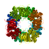
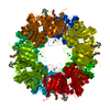
| Entry | Database: EMDB / ID: EMD-2864 | |||||||||
|---|---|---|---|---|---|---|---|---|---|---|
| Title | Vitrified IAS Env | |||||||||
 Map data Map data | Moloney mouse leukemia virus Env | |||||||||
 Sample Sample |
| |||||||||
 Keywords Keywords |  Cryo-electron microscopy / Env trimers / image processing / isomerization arrested state / receptor binding domain Cryo-electron microscopy / Env trimers / image processing / isomerization arrested state / receptor binding domain | |||||||||
| Biological species |  Moloney murine leukemia virus Moloney murine leukemia virus | |||||||||
| Method | single particle reconstruction / cryo EM / negative staining / Resolution: 19.0 Å | |||||||||
 Authors Authors | Wu S-R / Sjoberg M / Wallin M / Lindqvist B / Ekstrom M / Hebert H / Koeck P / Garoff H | |||||||||
 Citation Citation | Journal: EMBO J / Year: 2008 Title: Turning of the receptor-binding domains opens up the murine leukaemia virus Env for membrane fusion. Authors: Shang-Rung Wu / Mathilda Sjöberg / Michael Wallin / Birgitta Lindqvist / Maria Ekström / Hans Hebert / Philip J B Koeck / Henrik Garoff /  Abstract: The activity of the membrane fusion protein Env of Moloney mouse leukaemia virus is controlled by isomerization of the disulphide that couples its transmembrane (TM) and surface (SU) subunits. We ...The activity of the membrane fusion protein Env of Moloney mouse leukaemia virus is controlled by isomerization of the disulphide that couples its transmembrane (TM) and surface (SU) subunits. We have arrested Env activation at a stage prior to isomerization by alkylating the active thiol in SU and compared the structure of isomerization-arrested Env with that of native Env. Env trimers of respective form were isolated from solubilized particles by sedimentation and their structures were reconstructed from electron microscopic images of both vitrified and negatively stained samples. We found that the protomeric unit of both trimers formed three protrusions, a top, middle and a lower one. The atomic structure of the receptor-binding domain of SU fitted into the upper protrusion. This was formed similar to a bent finger. Significantly, in native Env the tips of the fingers were directed against each other enclosing a cavity below, whereas they had turned outward in isomerization-arrested Env transforming the cavity into an open well. This might subsequently guide the fusion peptides in extended TM subunits into the target membrane. | |||||||||
| History |
|
- Structure visualization
| Movie |
 Movie viewer Movie viewer |
|---|---|
| Structure viewer | EM map: SurfViewMolmilJmol/JSmol |
| Supplemental images |

- Downloads & links
-EMDB archive
| Map data | emd_2864.map.gz | 382.7 KB | EMDB map data format | |
|---|---|---|---|---|
| Header (meta data) | emd-2864-v30.xmlemd-2864.xml | 9.8 KB 9.8 KB | Display Display | EMDB header |
| Images | 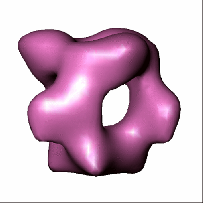 2864.gif 2864.gif | 45 KB | ||
| Others | cryo_IAS.map | 433 KB | ||
| Archive directory |  http://ftp.pdbj.org/pub/emdb/structures/EMD-2864ftp://ftp.pdbj.org/pub/emdb/structures/EMD-2864 http://ftp.pdbj.org/pub/emdb/structures/EMD-2864ftp://ftp.pdbj.org/pub/emdb/structures/EMD-2864 | HTTPS FTP |
-Related structure data

-Links
| EMDB pages | EMDB (EBI/PDBe) / EMDataResource |
|---|
-Map
| File | Download / File: emd_2864.map.gz / Format: CCP4 / Size: 422.9 KB / Type: IMAGE STORED AS FLOATING POINT NUMBER (4 BYTES) | ||||||||||||||||||||||||||||||||||||||||||||||||||||||||||||
|---|---|---|---|---|---|---|---|---|---|---|---|---|---|---|---|---|---|---|---|---|---|---|---|---|---|---|---|---|---|---|---|---|---|---|---|---|---|---|---|---|---|---|---|---|---|---|---|---|---|---|---|---|---|---|---|---|---|---|---|---|---|
| Annotation | Moloney mouse leukemia virus Env | ||||||||||||||||||||||||||||||||||||||||||||||||||||||||||||
| Voxel size | X=Y=Z: 3.5 Å | ||||||||||||||||||||||||||||||||||||||||||||||||||||||||||||
| Density |
| ||||||||||||||||||||||||||||||||||||||||||||||||||||||||||||
| Symmetry | Space group: 1 | ||||||||||||||||||||||||||||||||||||||||||||||||||||||||||||
| Details | EMDB XML:
CCP4 map header:
| ||||||||||||||||||||||||||||||||||||||||||||||||||||||||||||
-Supplemental data
-Supplemental map: cryo IAS.map
| File | cryo_IAS.map | ||||||||||||
|---|---|---|---|---|---|---|---|---|---|---|---|---|---|
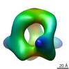
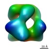
| Projections & Slices |
| ||||||||||||
| Density Histograms |
 Z
Z Y
Y X
X


- Sample components
-Entire : Moloney mouse leukemia virus Env
| Entire | Name: Moloney mouse leukemia virus Env |
|---|---|
| Components |
|
-Supramolecule #1000: Moloney mouse leukemia virus Env
| Supramolecule | Name: Moloney mouse leukemia virus Env / type: sample / ID: 1000 / Oligomeric state: Trimer / Number unique components: 1 |
|---|---|
| Molecular weight | Experimental: 200 KDa / Method: Gel electrophoresis |
-Macromolecule #1: Moloney murine leukemia virus
| Macromolecule | Name: Moloney murine leukemia virus / type: protein_or_peptide / ID: 1 / Name.synonym: Mo-MLV Env / Number of copies: 3 / Oligomeric state: Trimer / Recombinant expression: Yes |
|---|---|
| Source (natural) | Organism: Moloney murine leukemia virus / Tissue: Virus / Cell: MOV-3, NIH 3T3 |
| Molecular weight | Experimental: 200 KDa |
-Experimental details
-Structure determination
| Method | negative staining, cryo EM |
|---|---|
 Processing Processing | single particle reconstruction |
| Aggregation state | particle |
-Sample preparation
| Buffer | pH: 7.4 Details: 20 mM HEPES, 135 mM NaCl, pH 7.45, 10mM EDTA, 0.15 % Triton X-100, 12 % (w/w) sucrose |
|---|---|
| Staining | Type: NEGATIVE / Details: No staining |
| Grid | Details: 400 mesh grid |
| Vitrification | Cryogen name: ETHANE / Chamber humidity: 99 % / Chamber temperature: 77 K / Instrument: OTHER / Details: Vitrification instrument: Vitrobot / Method: Blot for 3 seconds twice before plunging |
- Electron microscopy
| Microscope | FEI TECNAI F20 |
|---|---|
| Electron beam | Acceleration voltage: 300 kV / Electron source: FIELD EMISSION GUN |
| Electron optics | Illumination mode: SPOT SCAN / Imaging mode: BRIGHT FIELDBright-field microscopy / Cs: 2 mm / Nominal defocus max: 3.5 µm / Nominal defocus min: 2.5 µm / Nominal magnification: 86000 |
| Sample stage | Specimen holder: Eucentric / Specimen holder model: GATAN LIQUID NITROGEN |
| Temperature | Min: 93 K / Max: 96 K / Average: 95 K |
| Alignment procedure | Legacy - Astigmatism: Objective lens astigmatism was corrected using online FFT |
| Date | May 10, 2008 |
| Image recording | Category: CCD / Film or detector model: GENERIC CCD / Digitization - Sampling interval: 30 µm / Number real images: 96 / Average electron dose: 9 e/Å2 / Bits/pixel: 8 |
| Tilt angle min | 0 |
| Tilt angle max | 0 |
| Experimental equipment |  Model: Tecnai F20 / Image courtesy: FEI Company |
-Image processing
| CTF correction | Details: Each particle |
|---|---|
| Final two d classification | Number classes: 131 |
| Final reconstruction | Applied symmetry - Point group: C3 (3 fold cyclic) / Algorithm: OTHER / Resolution.type: BY AUTHOR / Resolution: 19.0 Å / Resolution method: FSC 0.5 CUT-OFF / Software - Name: EMAN / Number images used: 6787 |
