 Movie
Movie Controller


+ Open data

- Basic information
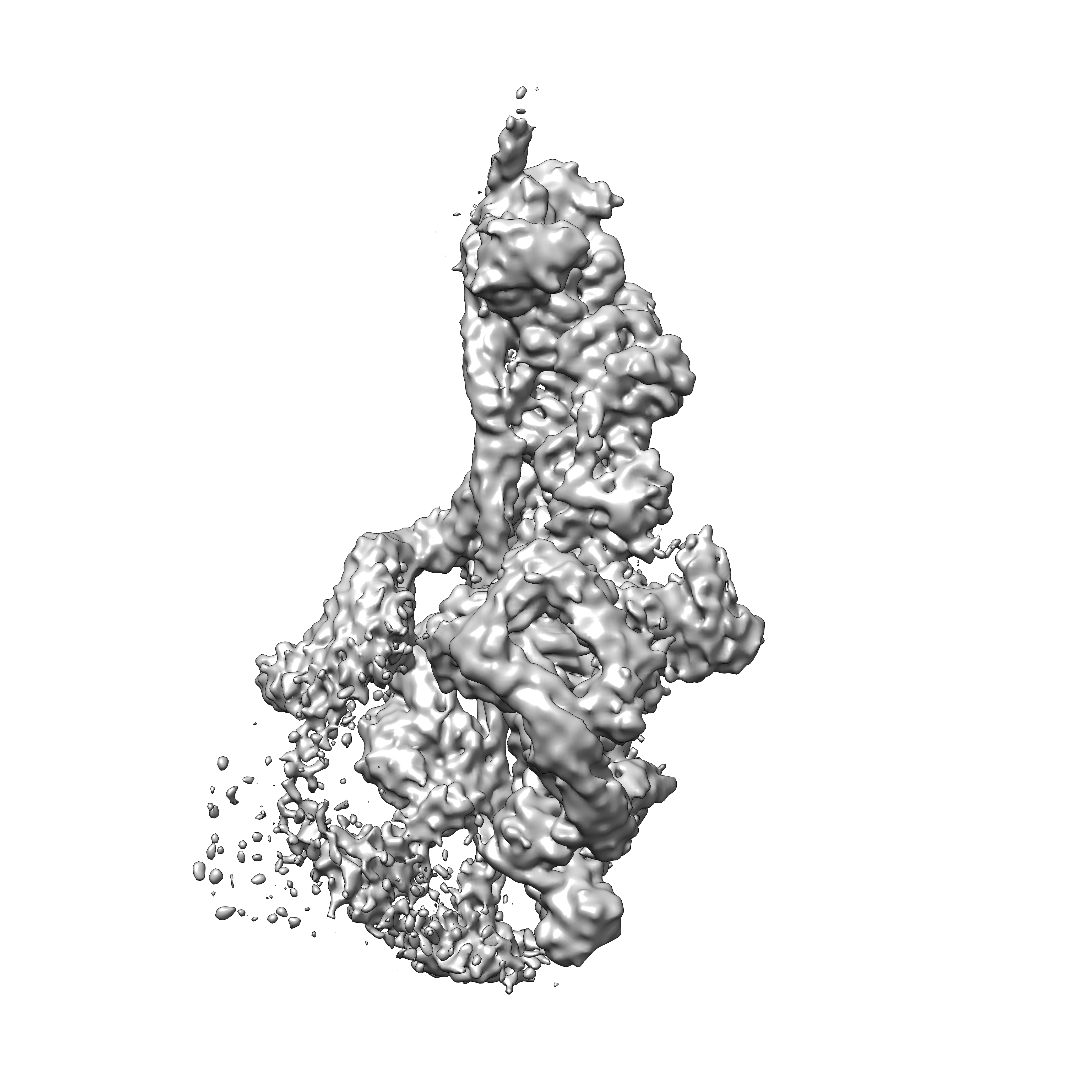
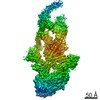
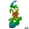
| Entry | Database: EMDB / ID: EMD-2860 | |||||||||
|---|---|---|---|---|---|---|---|---|---|---|
| Title | Electron cryo-microscopy of dynein/dynactin/GFP-BICD2N complex | |||||||||
 Map data Map data | Reconstruction of dynein tail/dynactin/GFP-BICD2N complex. | |||||||||
 Sample Sample |
| |||||||||
 Keywords Keywords |  dynein / dynactin / BICD2 / motor / transport dynein / dynactin / BICD2 / motor / transport | |||||||||
| Function / homology |  Function and homology information Function and homology informationretrograde axonal transport of mitochondrion / Gap junction degradation / Formation of annular gap junctions / Regulation of actin dynamics for phagocytic cup formation / EPHB-mediated forward signaling / VEGFA-VEGFR2 Pathway / Cell-extracellular matrix interactions / RHO GTPases Activate WASPs and WAVEs / MAP2K and MAPK activation / dynactin complex ...retrograde axonal transport of mitochondrion / Gap junction degradation / Formation of annular gap junctions / Regulation of actin dynamics for phagocytic cup formation / EPHB-mediated forward signaling / VEGFA-VEGFR2 Pathway / Cell-extracellular matrix interactions / RHO GTPases Activate WASPs and WAVEs / MAP2K and MAPK activation / dynactin complex / Clathrin-mediated endocytosis / transport along microtubule / visual behavior / dynein light chain binding / WASH complex / F-actin capping protein complex / dynein heavy chain binding / negative regulation of filopodium assembly / positive regulation of intracellular transport / regulation of metaphase plate congression / cellular response to cytochalasin B / establishment of spindle localization / ciliary tip / positive regulation of spindle assembly / regulation of transepithelial transport / structural constituent of postsynaptic actin cytoskeleton / morphogenesis of a polarized epithelium / Intraflagellar transport / postsynaptic actin cytoskeleton / protein localization to adherens junction / dense body / Tat protein binding / Neutrophil degranulation / P-body assembly / dynein complex / COPI-independent Golgi-to-ER retrograde traffic / apical protein localization / minus-end-directed microtubule motor activity / barbed-end actin filament capping / cytoplasmic dynein complex / retrograde axonal transport / adherens junction assembly / coronary vasculature development / dynein light intermediate chain binding / RHO GTPases activate IQGAPs / regulation of cell morphogenesis / RHO GTPases Activate Formins / regulation of lamellipodium assembly / HSP90 chaperone cycle for steroid hormone receptors (SHR) in the presence of ligand / COPI-independent Golgi-to-ER retrograde traffic / MHC class II antigen presentation / tight junction / nuclear migration / regulation of norepinephrine uptake / COPI-mediated anterograde transport / aorta development / NuA4 histone acetyltransferase complex / centrosome localization / regulation of synaptic vesicle endocytosis / ventricular septum development / microtubule motor activity / apical junction complex / establishment or maintenance of cell polarity / dynein intermediate chain binding / dynein complex binding / cortical cytoskeleton / positive regulation of double-strand break repair via homologous recombination / microtubule-based movement / nitric-oxide synthase binding / brush border / kinesin binding / calyx of Held / cytoplasmic microtubule / Amplification of signal from unattached kinetochores via a MAD2 inhibitory signal / regulation of protein localization to plasma membrane / microtubule-based process / COPI-mediated anterograde transport / stress granule assembly / cytoplasmic microtubule organization / stress fiber / Mitotic Prometaphase / cytoskeleton organization / regulation of mitotic spindle organization / EML4 and NUDC in mitotic spindle formation / axon cytoplasm / Loss of Nlp from mitotic centrosomes / Loss of proteins required for interphase microtubule organization from the centrosome / Recruitment of mitotic centrosome proteins and complexes / Resolution of Sister Chromatid Cohesion / Recruitment of NuMA to mitotic centrosomes / HSP90 chaperone cycle for steroid hormone receptors (SHR) in the presence of ligand / Anchoring of the basal body to the plasma membrane / MHC class II antigen presentation / axonogenesis / sarcomere / AURKA Activation by TPX2 / cellular response to nerve growth factor stimulus / mitotic spindle organization / filopodium / actin filamentSimilarity search - Function | |||||||||
| Biological species |  Homo sapiens (human) / Homo sapiens (human) /  Mus musculus (house mouse) / Sus scrofa domesticus (domestic pig) Mus musculus (house mouse) / Sus scrofa domesticus (domestic pig) | |||||||||
| Method | single particle reconstruction / cryo EM / Resolution: 8.2 Å | |||||||||
 Authors Authors | Urnavicius L / Zhang K / Diamant AG / Motz C / Schlager MA / Yu M / Patel NA / Robinson CV / Carter AP | |||||||||
 Citation Citation | Journal: Science / Year: 2015 Title: The structure of the dynactin complex and its interaction with dynein. Authors: Linas Urnavicius / Kai Zhang / Aristides G Diamant / Carina Motz / Max A Schlager / Minmin Yu / Nisha A Patel / Carol V Robinson / Andrew P Carter /  Abstract: Dynactin is an essential cofactor for the microtubule motor cytoplasmic dynein-1. We report the structure of the 23-subunit dynactin complex by cryo-electron microscopy to 4.0 angstroms. Our ...Dynactin is an essential cofactor for the microtubule motor cytoplasmic dynein-1. We report the structure of the 23-subunit dynactin complex by cryo-electron microscopy to 4.0 angstroms. Our reconstruction reveals how dynactin is built around a filament containing eight copies of the actin-related protein Arp1 and one of β-actin. The filament is capped at each end by distinct protein complexes, and its length is defined by elongated peptides that emerge from the α-helical shoulder domain. A further 8.2 angstrom structure of the complex between dynein, dynactin, and the motility-inducing cargo adaptor Bicaudal-D2 shows how the translational symmetry of the dynein tail matches that of the dynactin filament. The Bicaudal-D2 coiled coil runs between dynein and dynactin to stabilize the mutually dependent interactions between all three components. | |||||||||
| History |
|
- Structure visualization
| Movie |
Movie viewer |
|---|---|
| Structure viewer | EM map: SurfViewMolmilJmol/JSmol |
| Supplemental images |
- Downloads & links
-EMDB archive
| Map data | emd_2860.map.gz | 10.9 MB | EMDB map data format | |
|---|---|---|---|---|
| Header (meta data) | emd-2860-v30.xmlemd-2860.xml | 10.8 KB 10.8 KB | Display Display | EMDB header |
| Images |  emd_2860.png emd_2860.png | 2.2 MB | ||
| Archive directory |  http://ftp.pdbj.org/pub/emdb/structures/EMD-2860ftp://ftp.pdbj.org/pub/emdb/structures/EMD-2860 http://ftp.pdbj.org/pub/emdb/structures/EMD-2860ftp://ftp.pdbj.org/pub/emdb/structures/EMD-2860 | HTTPS FTP |
-Related structure data
| Related structure data |  5afuMC  6f3aM 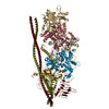 6zo4M 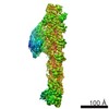 2854C 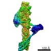 2855C 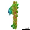 2856C 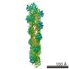 2857C 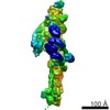 2861C 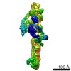 2862C  5adxC  5afrC M: atomic model generated by this map C: citing same article ( |
|---|---|
| Similar structure data |
-Links
| EMDB pages | EMDB (EBI/PDBe) / EMDataResource |
|---|---|
| Related items in Molecule of the Month |


-Map
| File | Download / File: emd_2860.map.gz / Format: CCP4 / Size: 300.3 MB / Type: IMAGE STORED AS FLOATING POINT NUMBER (4 BYTES) | ||||||||||||||||||||||||||||||||||||||||||||||||||||||||||||
|---|---|---|---|---|---|---|---|---|---|---|---|---|---|---|---|---|---|---|---|---|---|---|---|---|---|---|---|---|---|---|---|---|---|---|---|---|---|---|---|---|---|---|---|---|---|---|---|---|---|---|---|---|---|---|---|---|---|---|---|---|---|
| Annotation | Reconstruction of dynein tail/dynactin/GFP-BICD2N complex. | ||||||||||||||||||||||||||||||||||||||||||||||||||||||||||||
| Voxel size | X=Y=Z: 1.34 Å | ||||||||||||||||||||||||||||||||||||||||||||||||||||||||||||
| Density |
| ||||||||||||||||||||||||||||||||||||||||||||||||||||||||||||
| Symmetry | Space group: 1 | ||||||||||||||||||||||||||||||||||||||||||||||||||||||||||||
| Details | EMDB XML:
CCP4 map header:
| ||||||||||||||||||||||||||||||||||||||||||||||||||||||||||||
-Supplemental data
- Sample components
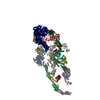
-Entire : Cryo-EM structure of dynein tail/dynactin/GFP-BICD2N
| Entire | Name: Cryo-EM structure of dynein tail/dynactin/GFP-BICD2N |
|---|---|
| Components |
|
-Supramolecule #1000: Cryo-EM structure of dynein tail/dynactin/GFP-BICD2N
| Supramolecule | Name: Cryo-EM structure of dynein tail/dynactin/GFP-BICD2N / type: sample / ID: 1000 / Details: The sample was monodisperse / Number unique components: 3 |
|---|---|
| Molecular weight | Theoretical: 2.6 MDa |
-Macromolecule #1: Human cytoplasmic dynein 1 tail
| Macromolecule | Name: Human cytoplasmic dynein 1 tail / type: protein_or_peptide / ID: 1 / Name.synonym: TDB / Number of copies: 1 / Recombinant expression: No |
|---|---|
| Source (natural) | Organism: Homo sapiens (human) / synonym: Human//Mouse / Location in cell: cytoplasm |
| Molecular weight | Theoretical: 2.6 MDa |
-Macromolecule #2: mouse BICD2N
| Macromolecule | Name: mouse BICD2N / type: protein_or_peptide / ID: 2 / Number of copies: 1 / Recombinant expression: No |
|---|---|
| Source (natural) | Organism: Mus musculus (house mouse) / synonym: house mouse |
-Macromolecule #3: pig dynactin
| Macromolecule | Name: pig dynactin / type: protein_or_peptide / ID: 3 / Number of copies: 1 / Recombinant expression: No |
|---|---|
| Source (natural) | Organism: Sus scrofa domesticus (domestic pig) / synonym: domestic pig / Location in cell: cytoplasm |
-Experimental details
-Structure determination
| Method | cryo EM |
|---|---|
 Processing Processing | single particle reconstruction |
| Aggregation state | particle |
-Sample preparation
| Concentration | 0.075 mg/mL |
|---|---|
| Buffer | pH: 7.4 Details: 150mM KCl, 25mM HEPES-KOH, 1mM MgCl2, 0.1mM MgATP, 5mM DTT |
| Grid | Details: Quantifoil R1.2/1.3 400 mesh copper grid with thin carbon support, glow discharged |
| Vitrification | Cryogen name: ETHANE / Chamber humidity: 100 % / Chamber temperature: 120 K / Instrument: FEI VITROBOT MARK III / Method: Blot for 3.5 seconds before plunging |
- Electron microscopy
| Microscope | FEI TITAN KRIOS |
|---|---|
| Electron beam | Acceleration voltage: 300 kV / Electron source: FIELD EMISSION GUN |
| Electron optics | Calibrated magnification: 82353 / Illumination mode: FLOOD BEAM / Imaging mode: BRIGHT FIELDBright-field microscopy / Cs: 2.7 mm / Nominal defocus max: 8.0 µm / Nominal defocus min: 3.0 µm / Nominal magnification: 47000 |
| Sample stage | Specimen holder model: FEI TITAN KRIOS AUTOGRID HOLDER |
| Temperature | Average: 100 K |
| Alignment procedure | Legacy - Astigmatism: Objective lens astigmatism was corrected at 120,000 times magnification. |
| Date | Jul 17, 2014 |
| Image recording | Category: CCD / Film or detector model: FEI FALCON II (4k x 4k) / Number real images: 4259 / Average electron dose: 1 e/Å2 / Bits/pixel: 16 |
| Experimental equipment |  Model: Titan Krios / Image courtesy: FEI Company |
-Image processing
| CTF correction | Details: Each particle |
|---|---|
| Final reconstruction | Applied symmetry - Point group: C1 (asymmetric) / Algorithm: OTHER / Resolution.type: BY AUTHOR / Resolution: 8.2 Å / Resolution method: OTHER / Software - Name: Relion / Number images used: 85744 |
| Details | The particles were selected using an automatic selection program. |
