 Movie
Movie Controller

[English] 日本語
 Yorodumi
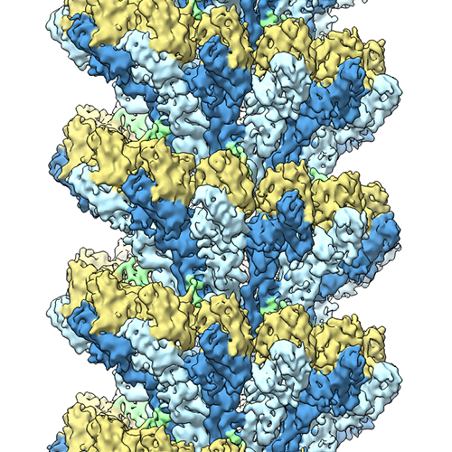
- EMDB-2799: Cryo-EM structure of gamma-TuSC oligomers in a closed conformation -
+ Open data

- Basic information
| Entry | Database: EMDB / ID: EMD-2799 | |||||||||
|---|---|---|---|---|---|---|---|---|---|---|
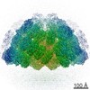
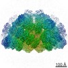
| Title | Cryo-EM structure of gamma-TuSC oligomers in a closed conformation | |||||||||
 Map data Map data | Reconstruction of yeast gamma-TuSC trapped in a closed state by disulfide crosslinks | |||||||||
 Sample Sample |
| |||||||||
 Keywords Keywords |  Microtubule nucleation / gamma tubulin Microtubule nucleation / gamma tubulin | |||||||||
| Function / homology |  Function and homology information Function and homology informationgamma-tubulin complex localization to nuclear side of mitotic spindle pole body / protein localization to mitotic spindle pole body / inner plaque of spindle pole body / microtubule nucleation by spindle pole body / outer plaque of spindle pole body / central plaque of spindle pole body / gamma-tubulin small complex / regulation of microtubule nucleation / karyogamy involved in conjugation with cellular fusion / mitotic spindle pole body ...gamma-tubulin complex localization to nuclear side of mitotic spindle pole body / protein localization to mitotic spindle pole body / inner plaque of spindle pole body / microtubule nucleation by spindle pole body / outer plaque of spindle pole body / central plaque of spindle pole body / gamma-tubulin small complex / regulation of microtubule nucleation / karyogamy involved in conjugation with cellular fusion / mitotic spindle pole body / equatorial microtubule organizing center / mitotic spindle elongation / gamma-tubulin complex / meiotic spindle organization / positive regulation of microtubule nucleation / microtubule nucleation / positive regulation of cytoplasmic translation / spindle pole body / gamma-tubulin binding / mitotic sister chromatid segregation / spindle assembly / cytoplasmic microtubule organization / mitotic spindle organization / meiotic cell cycle / structural constituent of cytoskeleton / spindle / spindle pole / mitotic cell cycle / protein-containing complex assembly / microtubule / calmodulin binding / protein-containing complex binding / GTP binding / nucleus / cytoplasmSimilarity search - Function | |||||||||
| Biological species |  Saccharomyces cerevisiae (brewer's yeast) Saccharomyces cerevisiae (brewer's yeast) | |||||||||
| Method | helical reconstruction / cryo EM / Resolution: 6.9 Å | |||||||||
 Authors Authors | Kollman JM / Greenberg CH / Li S / Moritz M / Zelter A / Fong K / Fernandez J-J / Sali A / Kilmartin J / Davis TN / Agard DA | |||||||||
 Citation Citation | Journal: Nat Struct Mol Biol / Year: 2015 Title: Ring closure activates yeast γTuRC for species-specific microtubule nucleation. Authors: Justin M Kollman / Charles H Greenberg / Sam Li / Michelle Moritz / Alex Zelter / Kimberly K Fong / Jose-Jesus Fernandez / Andrej Sali / John Kilmartin / Trisha N Davis / David A Agard /    Abstract: The γ-tubulin ring complex (γTuRC) is the primary microtubule nucleator in cells. γTuRC is assembled from repeating γ-tubulin small complex (γTuSC) subunits and is thought to function as a ...The γ-tubulin ring complex (γTuRC) is the primary microtubule nucleator in cells. γTuRC is assembled from repeating γ-tubulin small complex (γTuSC) subunits and is thought to function as a template by presenting a γ-tubulin ring that mimics microtubule geometry. However, a previous yeast γTuRC structure showed γTuSC in an open conformation that prevents matching to microtubule symmetry. By contrast, we show here that γ-tubulin complexes are in a closed conformation when attached to microtubules. To confirm the functional importance of the closed γTuSC ring, we trapped the closed state and determined its structure, showing that the γ-tubulin ring precisely matches microtubule symmetry and providing detailed insight into γTuRC architecture. Importantly, the closed state is a stronger nucleator, thus suggesting that this conformational switch may allosterically control γTuRC activity. Finally, we demonstrate that γTuRCs have a strong preference for tubulin from the same species. | |||||||||
| History |
|
- Structure visualization
| Movie |
Movie viewer |
|---|---|
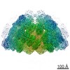
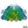
| Structure viewer | EM map: SurfViewMolmilJmol/JSmol |
| Supplemental images |
- Downloads & links
-EMDB archive
| Map data | emd_2799.map.gz | 33.3 MB | EMDB map data format | |
|---|---|---|---|---|
| Header (meta data) | emd-2799-v30.xmlemd-2799.xml | 13.7 KB 13.7 KB | Display Display | EMDB header |
| Images |  emd-2799_mapimage.png emd-2799_mapimage.png | 418.5 KB | ||
| Archive directory |  http://ftp.pdbj.org/pub/emdb/structures/EMD-2799ftp://ftp.pdbj.org/pub/emdb/structures/EMD-2799 http://ftp.pdbj.org/pub/emdb/structures/EMD-2799ftp://ftp.pdbj.org/pub/emdb/structures/EMD-2799 | HTTPS FTP |
-Related structure data
| Related structure data | 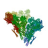 5flzM  5989C M: atomic model generated by this map C: citing same article ( |
|---|---|
| Similar structure data |

-Links
| EMDB pages | EMDB (EBI/PDBe) / EMDataResource |
|---|---|
| Related items in Molecule of the Month |


-Map
| File | Download / File: emd_2799.map.gz / Format: CCP4 / Size: 35.5 MB / Type: IMAGE STORED AS FLOATING POINT NUMBER (4 BYTES) | ||||||||||||||||||||||||||||||||||||||||||||||||||||||||||||
|---|---|---|---|---|---|---|---|---|---|---|---|---|---|---|---|---|---|---|---|---|---|---|---|---|---|---|---|---|---|---|---|---|---|---|---|---|---|---|---|---|---|---|---|---|---|---|---|---|---|---|---|---|---|---|---|---|---|---|---|---|---|
| Annotation | Reconstruction of yeast gamma-TuSC trapped in a closed state by disulfide crosslinks | ||||||||||||||||||||||||||||||||||||||||||||||||||||||||||||
| Voxel size | X=Y=Z: 1.88 Å | ||||||||||||||||||||||||||||||||||||||||||||||||||||||||||||
| Density |
| ||||||||||||||||||||||||||||||||||||||||||||||||||||||||||||
| Symmetry | Space group: 1 | ||||||||||||||||||||||||||||||||||||||||||||||||||||||||||||
| Details | EMDB XML:
CCP4 map header:
| ||||||||||||||||||||||||||||||||||||||||||||||||||||||||||||
-Supplemental data
- Sample components
-Entire : Recombinant yeast gamma-TuSC mutant S58C/G288C
| Entire | Name: Recombinant yeast gamma-TuSC mutant S58C/G288C |
|---|---|
| Components |
|
-Supramolecule #1000: Recombinant yeast gamma-TuSC mutant S58C/G288C
| Supramolecule | Name: Recombinant yeast gamma-TuSC mutant S58C/G288C / type: sample / ID: 1000 / Oligomeric state: heteropentamer / Number unique components: 4 |
|---|
-Macromolecule #1: gamma tubulin S58C/G288C
| Macromolecule | Name: gamma tubulin S58C/G288C / type: protein_or_peptide / ID: 1 / Name.synonym: tub4 Details: Cysteine residues were introduced at positions 58 and 288 to promote crosslinking of the helical complex. Number of copies: 2 / Recombinant expression: Yes |
|---|---|
| Source (natural) | Organism: Saccharomyces cerevisiae (brewer's yeast) / synonym: Baker's yeast / Location in cell: spindle pole body |
| Molecular weight | Theoretical: 55 KDa |
| Recombinant expression | Organism:  unidentified baculovirus unidentified baculovirus |
| Sequence | UniProtKB: Tubulin gamma chain |
-Macromolecule #2: GCP2
| Macromolecule | Name: GCP2 / type: protein_or_peptide / ID: 2 / Name.synonym: Spc97 / Number of copies: 1 / Recombinant expression: Yes |
|---|---|
| Source (natural) | Organism: Saccharomyces cerevisiae (brewer's yeast) / synonym: Baker's yeast / Location in cell: spindle pole body |
| Molecular weight | Theoretical: 97 KDa |
| Recombinant expression | Organism: unidentified baculovirus |
| Sequence | UniProtKB: Spindle pole body component SPC97 |
-Macromolecule #3: GCP3
| Macromolecule | Name: GCP3 / type: protein_or_peptide / ID: 3 / Name.synonym: Spc98 / Number of copies: 1 / Recombinant expression: Yes |
|---|---|
| Source (natural) | Organism: Saccharomyces cerevisiae (brewer's yeast) / synonym: Baker's yeast / Location in cell: spindle pole body |
| Molecular weight | Theoretical: 98 KDa |
| Recombinant expression | Organism: unidentified baculovirus |
| Sequence | UniProtKB: Spindle pole body component SPC98 |
-Macromolecule #4: Spc110 (1-220)
| Macromolecule | Name: Spc110 (1-220) / type: protein_or_peptide / ID: 4 Details: Residues 1-220 of Spc110 were expressed with an N-terminal GST tagged, which was cleaved off during purification. Number of copies: 2 / Oligomeric state: dimer / Recombinant expression: Yes |
|---|---|
| Source (natural) | Organism: Saccharomyces cerevisiae (brewer's yeast) / synonym: Baker's yeast / Location in cell: spindle pole body |
| Molecular weight | Theoretical: 25 KDa |
| Recombinant expression | Organism: unidentified baculovirus |
| Sequence | UniProtKB: Spindle pole body component 110 |
-Experimental details
-Structure determination
| Method | cryo EM |
|---|---|
 Processing Processing | helical reconstruction |
| Aggregation state | filament |
-Sample preparation
| Concentration | 2 mg/mL |
|---|---|
| Buffer | pH: 7.6 Details: 40 mM Hepes PH 7.6, 100 mM KCl, 1 mM EGTA, 1mM MgCl2, 1 mM oxidized glutathione |
| Grid | Details: 400 mesh C-FLAT 2/2 grid |
| Vitrification | Cryogen name: ETHANE / Chamber humidity: 90 % / Instrument: FEI VITROBOT MARK I / Method: Blot for 2-5 seconds before plunging |
- Electron microscopy
| Microscope | FEI TECNAI F20 |
|---|---|
| Electron beam | Acceleration voltage: 200 kV / Electron source: FIELD EMISSION GUN |
| Electron optics | Calibrated magnification: 94000 / Illumination mode: FLOOD BEAM / Imaging mode: BRIGHT FIELDBright-field microscopy / Cs: 2.12 mm / Nominal defocus max: 2.0 µm / Nominal defocus min: 0.8 µm |
| Sample stage | Specimen holder model: GATAN LIQUID NITROGEN |
| Alignment procedure | Legacy - Astigmatism: Objective astigmatism corrected at 135,000 time magnifiaction |
| Date | May 25, 2011 |
| Image recording | Category: CCD / Film or detector model: TVIPS TEMCAM-F816 (8k x 8k) / Number real images: 364 / Average electron dose: 20 e/Å2 |
| Experimental equipment |  Model: Tecnai F20 / Image courtesy: FEI Company |
-Image processing
| CTF correction | Details: each micrograph |
|---|---|
| Final reconstruction | Applied symmetry - Helical parameters - Δz: 22.2 Å Applied symmetry - Helical parameters - Δ&Phi: 54.3 ° Applied symmetry - Helical parameters - Axial symmetry: C1 (asymmetric) Algorithm: OTHER / Resolution.type: BY AUTHOR / Resolution: 6.9 Å / Resolution method: OTHER / Software - Name: SPIDER, hsearch_lorentz, EMAN1, ctffind |
| Details | IHRSR was carried out in SPIDER, using hsearch_lorentz to search for helical symmetry parameters in unsymmetrized maps. |
