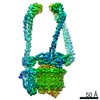
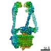
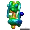
Journal: Nat Struct Mol Biol / Year: 2014 Title: Structure and mechanism of action of the BRCA2 breast cancer tumor suppressor. Authors: Taha Shahid / Joanna Soroka / Eric Kong / Laurent Malivert / Michael J McIlwraith / Tillman Pape / Stephen C West / Xiaodong Zhang / Abstract: Mutations in BRCA2 increase susceptibility to breast, ovarian and prostate cancers. The product of human BRCA2, BRCA2 protein, has a key role in the repair of DNA double-strand breaks and interstrand ...Mutations in BRCA2 increase susceptibility to breast, ovarian and prostate cancers. The product of human BRCA2, BRCA2 protein, has a key role in the repair of DNA double-strand breaks and interstrand cross-links by RAD51-mediated homologous recombination. Here, we present a biochemical and structural characterization of full-length (3,418 amino acid) BRCA2, alone and in complex with RAD51. We show that BRCA2 facilitates nucleation of RAD51 filaments at multiple sites on single-stranded DNA. Three-dimensional EM reconstructions revealed that BRCA2 exists as a dimer and that two oppositely oriented sets of RAD51 molecules bind the dimer. Single-stranded DNA binds along the long axis of BRCA2, such that only one set of RAD51 monomers can form a productive complex with DNA and establish filament formation. Our data define the molecular mechanism by which this tumor suppressor facilitates RAD51-mediated homologous-recombinational repair.
History
Deposition
Sep 3, 2014
-
Header (metadata) release
Sep 17, 2014
-
Map release
Oct 8, 2014
-
Update
Oct 8, 2014
-
Current status
Oct 8, 2014
Processing site: PDBe / Status: Released
-
Structure visualization
Movie
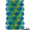
Surface view with section colored by density value
Macromolecule #1: Breast cancer type 2 susceptibility protein
Macromolecule
Name: Breast cancer type 2 susceptibility protein / type: protein_or_peptide / ID: 1 / Name.synonym: BRCA2 / Number of copies: 2 / Oligomeric state: Dimer / Recombinant expression: Yes
Source (natural)
Organism: Homo sapiens (human) / synonym: Human / Location in cell: Nucleus
Molecular weight
Theoretical: 384 KDa
Recombinant expression
Organism: Homo sapiens (human) / Recombinant cell: HeLa
Sequence
UniProtKB: Breast cancer type 2 susceptibility protein / InterPro: Breast cancer type 2 susceptibility protein
-
Macromolecule #2: DNA repair protein RAD51 homolog 1
Macromolecule
Name: DNA repair protein RAD51 homolog 1 / type: protein_or_peptide / ID: 2 / Name.synonym: HsRAD51 / Number of copies: 8 / Oligomeric state: Dimer of tetramers / Recombinant expression: Yes
Source (natural)
Organism: Homo sapiens (human) / synonym: Human / Location in cell: Nucleus
Molecular weight
Theoretical: 37 KDa
Recombinant expression
Organism: Escherichia coli (E. coli)
Sequence
UniProtKB: DNA repair protein RAD51 homolog 1
-
Experimental details
-
Structure determination
Method
negative staining
Processing
single particle reconstruction
Aggregation state
particle
-
Sample preparation
Staining
Type: NEGATIVE Details: Grid with adsorbed protein was washed twice with water and negatively stained with 2% uranyl acetate solution before being blotted and air dried.
Grid
Details: Copper Quantifoil R2/2 holey carbon grids coated with continuous carbon, glow discharged in air.
Vitrification
Cryogen name: NONE / Instrument: OTHER
-
Electron microscopy #1
Microscopy ID
1
Microscope
FEI/PHILIPS CM200FEG
Alignment procedure
Legacy - Astigmatism: Objective lens astigmatism was corrected at 150,000 times magnification.
Date
Dec 14, 2012
Image recording
Category: CCD / Film or detector model: TVIPS TEMCAM-F415 (4k x 4k) / Average electron dose: 40 e/Å2 / Bits/pixel: 16
Electron beam
Acceleration voltage: 200 kV / Electron source: FIELD EMISSION GUN
Applied symmetry - Point group: C2 (2 fold cyclic) / Algorithm: OTHER / Resolution.type: BY AUTHOR / Resolution: 19.5 Å / Resolution method: OTHER / Software - Name: IMAGIC-5, Tigris Details: Final maps were calculated after a round of projection matching. Number images used: 6877
In the structure databanks used in Yorodumi, some data are registered as the other names, "COVID-19 virus" and "2019-nCoV". Here are the details of the virus and the list of structure data.
Jan 31, 2019. EMDB accession codes are about to change! (news from PDBe EMDB page)
EMDB accession codes are about to change! (news from PDBe EMDB page)
The allocation of 4 digits for EMDB accession codes will soon come to an end. Whilst these codes will remain in use, new EMDB accession codes will include an additional digit and will expand incrementally as the available range of codes is exhausted. The current 4-digit format prefixed with “EMD-” (i.e. EMD-XXXX) will advance to a 5-digit format (i.e. EMD-XXXXX), and so on. It is currently estimated that the 4-digit codes will be depleted around Spring 2019, at which point the 5-digit format will come into force.
The EM Navigator/Yorodumi systems omit the EMD- prefix.
Related info.:Q: What is EMD? / ID/Accession-code notation in Yorodumi/EM Navigator
Yorodumi is a browser for structure data from EMDB, PDB, SASBDB, etc.
This page is also the successor to EM Navigator detail page, and also detail information page/front-end page for Omokage search.
The word "yorodu" (or yorozu) is an old Japanese word meaning "ten thousand". "mi" (miru) is to see.
Related info.:EMDB / PDB / SASBDB / Comparison of 3 databanks / Yorodumi Search / Aug 31, 2016. New EM Navigator & Yorodumi / Yorodumi Papers / Jmol/JSmol / Function and homology information / Changes in new EM Navigator and Yorodumi
 Movie
Movie Controller

 Yorodumi
 Open data
 Basic information
 Map data
 Sample
 Keywords
 Function and homology information
 Homo sapiens (human)
 Authors
 Citation
 Structure visualization

 Downloads & links
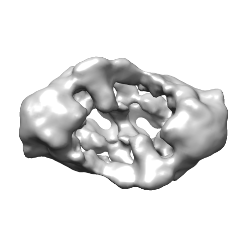 emd_2780.png
 emd_2780_1.png
 http://ftp.pdbj.org/pub/emdb/structures/EMD-2780
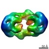


 Z (Sec.)
 Y (Row.)
 X (Col.)


 Sample components
 Processing
 Electron microscopy #1
 FIELD EMISSION GUN

