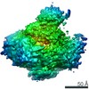
Movie
Movie Controller


+ Open data

- Basic information
| Entry | Database: EMDB / ID: EMD-2714 | |||||||||
|---|---|---|---|---|---|---|---|---|---|---|
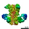
| Title | Negative stain reconstruction of AcrB/SMALP complex | |||||||||
 Map data Map data | Reconstruction of AcrB encapsulated in a SMALP polymer | |||||||||
 Sample Sample |
| |||||||||
 Keywords Keywords |  AcrB / negative stain / SMALP AcrB / negative stain / SMALP | |||||||||
| Function / homology |  Function and homology information Function and homology informationxenobiotic detoxification by transmembrane export across the cell outer membrane / efflux pump complex / periplasmic side of plasma membrane / xenobiotic transmembrane transporter activity / efflux transmembrane transporter activity / outer membrane-bounded periplasmic space / membrane / identical protein binding / plasma membraneSimilarity search - Function | |||||||||
| Biological species |  Escherichia coli (E. coli) Escherichia coli (E. coli) | |||||||||
| Method | single particle reconstruction / negative staining / Resolution: 23.0 Å | |||||||||
 Authors Authors | Postis V / Rawson S / Mitchell JK / Lee SC / Parslow R / Dafforn TR / Baldwin SA / Muench SP | |||||||||
 Citation Citation | Journal: Biochim Biophys Acta / Year: 2015 Title: The use of SMALPs as a novel membrane protein scaffold for structure study by negative stain electron microscopy. Authors: Vincent Postis / Shaun Rawson / Jennifer K Mitchell / Sarah C Lee / Rosemary A Parslow / Tim R Dafforn / Stephen A Baldwin / Stephen P Muench /  Abstract: Despite the great progress recently made in resolving their structures, investigation of the structural biology of membrane proteins still presents major challenges. Even with new technical advances ...Despite the great progress recently made in resolving their structures, investigation of the structural biology of membrane proteins still presents major challenges. Even with new technical advances such as lipidic cubic phase crystallisation, obtaining well-ordered crystals remains a significant hurdle in membrane protein X-ray crystallographic studies. As an alternative, electron microscopy has been shown to be capable of resolving >3.5Å resolution detail in membrane proteins of modest (~300 kDa) size, without the need for crystals. However, the conventional use of detergents for either approach presents several issues, including the possible effects on structure of removing the proteins from their natural membrane environment. As an alternative, it has recently been demonstrated that membrane proteins can be effectively isolated, in the absence of detergents, using a styrene maleic acid co-polymer (SMA). This approach yields SMA lipid particles (SMALPs) in which the membrane proteins are surrounded by a small disk of lipid bilayer encircled by polymer. Here we use the Escherichia coli secondary transporter AcrB as a model membrane protein to demonstrate how a SMALP scaffold can be used to visualise membrane proteins, embedded in a near-native lipid environment, by negative stain electron microscopy, yielding structures at a modest resolution in a short (days) timeframe. Moreover, we show that AcrB within a SMALP scaffold is significantly more active than the equivalent DDM stabilised form. The advantages of SMALP scaffolds within electron microscopy are discussed and we conclude that they may prove to be an important tool in studying membrane protein structure and function. | |||||||||
| History |
|
- Structure visualization
| Movie |
Movie viewer |
|---|---|
| Structure viewer | EM map: SurfViewMolmilJmol/JSmol |
| Supplemental images |

- Downloads & links
-EMDB archive
| Map data | emd_2714.map.gz | 6 MB | EMDB map data format | |
|---|---|---|---|---|
| Header (meta data) | emd-2714-v30.xmlemd-2714.xml | 10.1 KB 10.1 KB | Display Display | EMDB header |
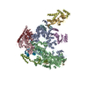
| Images | 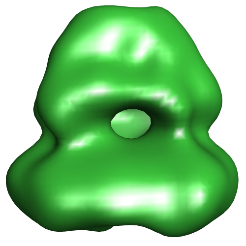 EMD-2714.png EMD-2714.png | 92.1 KB | ||
| Archive directory |  http://ftp.pdbj.org/pub/emdb/structures/EMD-2714ftp://ftp.pdbj.org/pub/emdb/structures/EMD-2714 http://ftp.pdbj.org/pub/emdb/structures/EMD-2714ftp://ftp.pdbj.org/pub/emdb/structures/EMD-2714 | HTTPS FTP |
-Related structure data
| Similar structure data |
|---|
-Links
| EMDB pages | EMDB (EBI/PDBe) / EMDataResource |
|---|---|
| Related items in Molecule of the Month |

-Map
| File | Download / File: emd_2714.map.gz / Format: CCP4 / Size: 7.8 MB / Type: IMAGE STORED AS FLOATING POINT NUMBER (4 BYTES) | ||||||||||||||||||||||||||||||||||||||||||||||||||||||||||||||||||||
|---|---|---|---|---|---|---|---|---|---|---|---|---|---|---|---|---|---|---|---|---|---|---|---|---|---|---|---|---|---|---|---|---|---|---|---|---|---|---|---|---|---|---|---|---|---|---|---|---|---|---|---|---|---|---|---|---|---|---|---|---|---|---|---|---|---|---|---|---|---|
| Annotation | Reconstruction of AcrB encapsulated in a SMALP polymer | ||||||||||||||||||||||||||||||||||||||||||||||||||||||||||||||||||||
| Voxel size | X=Y=Z: 4 Å | ||||||||||||||||||||||||||||||||||||||||||||||||||||||||||||||||||||
| Density |
| ||||||||||||||||||||||||||||||||||||||||||||||||||||||||||||||||||||
| Symmetry | Space group: 1 | ||||||||||||||||||||||||||||||||||||||||||||||||||||||||||||||||||||
| Details | EMDB XML:
CCP4 map header:
| ||||||||||||||||||||||||||||||||||||||||||||||||||||||||||||||||||||
-Supplemental data
- Sample components
-Entire : E. coli AcrB in a SMALP scaffold
| Entire | Name: E. coli AcrB in a SMALP scaffold |
|---|---|
| Components |
|
-Supramolecule #1000: E. coli AcrB in a SMALP scaffold
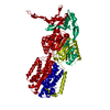
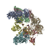
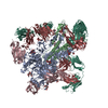
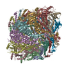
| Supramolecule | Name: E. coli AcrB in a SMALP scaffold / type: sample / ID: 1000 / Details: The sample was monodisperse / Oligomeric state: Trimeric AcrB / Number unique components: 1 |
|---|---|
| Molecular weight | Experimental: 360 KDa / Theoretical: 360 KDa |
-Macromolecule #1: Acridine resistance protein B
| Macromolecule | Name: Acridine resistance protein B / type: protein_or_peptide / ID: 1 / Name.synonym: AcrB / Number of copies: 3 / Oligomeric state: Trimer / Recombinant expression: Yes |
|---|---|
| Source (natural) | Organism: Escherichia coli (E. coli) / synonym: E. coli / Location in cell: membrane |
| Molecular weight | Experimental: 360 KDa / Theoretical: 360 KDa |
| Recombinant expression | Organism: Escherichia coli (E. coli) |
| Sequence | UniProtKB: Multidrug efflux pump subunit AcrB |
-Experimental details
-Structure determination
| Method | negative staining |
|---|---|
 Processing Processing | single particle reconstruction |
| Aggregation state | particle |
-Sample preparation
| Concentration | 0.12 mg/mL |
|---|---|
| Buffer | pH: 8 Details: 50mM Tris-HCl, pH 8.0, 10%(v/v) glycerol and 500mM NaCl |
| Staining | Type: NEGATIVE / Details: 1% Uranyl acetate |
| Grid | Details: Agar Carbon support film 300 mesh |
| Vitrification | Cryogen name: NONE / Instrument: OTHER |
- Electron microscopy
| Microscope | FEI TECNAI 12 |
|---|---|
| Electron beam | Acceleration voltage: 120 kV / Electron source: LAB6 |
| Electron optics | Calibrated magnification: 37500 / Illumination mode: FLOOD BEAM / Imaging mode: BRIGHT FIELDBright-field microscopy / Cs: 6.3 mm / Nominal defocus max: 1.2 µm / Nominal defocus min: 0.6 µm / Nominal magnification: 30000 |
| Sample stage | Specimen holder model: OTHER |
| Alignment procedure | Legacy - Astigmatism: Objective lens astigmatism was corrected at 80,000 times magnification |
| Details | Low Dose |
| Date | Feb 6, 2014 |
| Image recording | Category: CCD / Film or detector model: GATAN ORIUS SC200 (2k x 2k) / Number real images: 307 / Average electron dose: 50 e/Å2 |
-Image processing
| CTF correction | Details: Each micrograph CTF-find 3 |
|---|---|
| Final reconstruction | Applied symmetry - Point group: C3 (3 fold cyclic) / Algorithm: OTHER / Resolution.type: BY AUTHOR / Resolution: 23.0 Å / Resolution method: OTHER / Software - Name: RELION Details: Gold standard FSC used in RELION for resolution determination. A simple elipsoid used as a starting model Number images used: 6884 |
| Details | Particles were picked manually using EMAN2 boxer program |
-Atomic model buiding 1
| Initial model | PDB ID: Chain - #0 - Chain ID: A / Chain - #1 - Chain ID: B / Chain - #2 - Chain ID: C |
|---|---|
| Software | Name: Chmiera |
| Details | The crystal structure was fitted as a rigid body trimer |
| Refinement | Space: REAL / Protocol: RIGID BODY FIT |