 Movie
Movie Controller


+ Open data

- Basic information
| Entry | Database: EMDB / ID: EMD-2699 | |||||||||
|---|---|---|---|---|---|---|---|---|---|---|
| Title | VipA/VipB, contractile sheath of the type VI secretion system Type VI secretion system Type VI secretion system | |||||||||
 Map data Map data | Structure of the native helical assembly isolated from Vibrio cholerae | |||||||||
 Sample Sample |
| |||||||||
 Keywords Keywords | VipA / VipB / Vibrio / T6SS / cryo-EM / sheath | |||||||||
| Function / homology | Type VI secretion system TssC-like / TssC1, N-terminal / TssC1, C-terminal / EvpB/VC_A0108, tail sheath N-terminal domain / EvpB/VC_A0108, tail sheath gpW/gp25-like domain / Type VI secretion system sheath protein TssB1 / Type VI secretion system, VipA, VC_A0107 or Hcp2 / Type VI secretion system contractile sheath large subunit / Type VI secretion system contractile sheath small subunit Function and homology information Function and homology information | |||||||||
| Biological species |  Vibrio cholerae O1 biovar El Tor str. N16961 (bacteria) Vibrio cholerae O1 biovar El Tor str. N16961 (bacteria) | |||||||||
| Method | helical reconstruction / cryo EM / Resolution: 3.5 Å | |||||||||
 Authors Authors | Kudryashev M / Wang R / Brackmann M / Scherer S / Maier T / DiMaio F / Baker D / Stahlberg H / Egelman EH / Basler M | |||||||||
 Citation Citation | Journal: Cell / Year: 2015 Title: Structure of the type VI secretion system contractile sheath. Authors: Mikhail Kudryashev / Ray Yu-Ruei Wang / Maximilian Brackmann / Sebastian Scherer / Timm Maier / David Baker / Frank DiMaio / Henning Stahlberg / Edward H Egelman / Marek Basler /   Abstract: Bacteria use rapid contraction of a long sheath of the type VI secretion system (T6SS) to deliver effectors into a target cell. Here, we present an atomic-resolution structure of a native contracted ...Bacteria use rapid contraction of a long sheath of the type VI secretion system (T6SS) to deliver effectors into a target cell. Here, we present an atomic-resolution structure of a native contracted Vibrio cholerae sheath determined by cryo-electron microscopy. The sheath subunits, composed of tightly interacting proteins VipA and VipB, assemble into a six-start helix. The helix is stabilized by a core domain assembled from four β strands donated by one VipA and two VipB molecules. The fold of inner and middle layers is conserved between T6SS and phage sheaths. However, the structure of the outer layer is distinct and suggests a mechanism of interaction of the bacterial sheath with an accessory ATPase, ClpV, that facilitates multiple rounds of effector delivery. Our results provide a mechanistic insight into assembly of contractile nanomachines that bacteria and phages use to translocate macromolecules across membranes. | |||||||||
| History |
|
- Structure visualization
| Movie |
Movie viewer |
|---|---|
| Structure viewer | EM map: SurfViewMolmilJmol/JSmol |
| Supplemental images |

- Downloads & links
-EMDB archive
| Map data | emd_2699.map.gz | 72.4 MB | EMDB map data format | |
|---|---|---|---|---|
| Header (meta data) | emd-2699-v30.xmlemd-2699.xml | 11.4 KB 11.4 KB | Display Display | EMDB header |
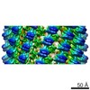
| Images | 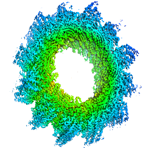 image2699.png image2699.png | 328.3 KB | ||
| Archive directory |  http://ftp.pdbj.org/pub/emdb/structures/EMD-2699ftp://ftp.pdbj.org/pub/emdb/structures/EMD-2699 http://ftp.pdbj.org/pub/emdb/structures/EMD-2699ftp://ftp.pdbj.org/pub/emdb/structures/EMD-2699 | HTTPS FTP |
-Related structure data
| Related structure data |  3j9gMC M: atomic model generated by this map C: citing same article ( |
|---|---|
| Similar structure data | |
| EM raw data | EMPIAR-10019 (Title: VipA/VipB, sheath of the bacterial type IV secretion system, micrographs for helical reconstruction taken on a K2 detector Data size: 4.1 Data #1: Frame-averaged micrographs [micrographs - single frame]) |


-Links
| EMDB pages | EMDB (EBI/PDBe) / EMDataResource |
|---|
-Map
| File | Download / File: emd_2699.map.gz / Format: CCP4 / Size: 268.2 MB / Type: IMAGE STORED AS FLOATING POINT NUMBER (4 BYTES) | ||||||||||||||||||||||||||||||||||||||||||||||||||||||||||||||||||||
|---|---|---|---|---|---|---|---|---|---|---|---|---|---|---|---|---|---|---|---|---|---|---|---|---|---|---|---|---|---|---|---|---|---|---|---|---|---|---|---|---|---|---|---|---|---|---|---|---|---|---|---|---|---|---|---|---|---|---|---|---|---|---|---|---|---|---|---|---|---|
| Annotation | Structure of the native helical assembly isolated from Vibrio cholerae | ||||||||||||||||||||||||||||||||||||||||||||||||||||||||||||||||||||
| Voxel size | X=Y=Z: 0.5 Å | ||||||||||||||||||||||||||||||||||||||||||||||||||||||||||||||||||||
| Density |
| ||||||||||||||||||||||||||||||||||||||||||||||||||||||||||||||||||||
| Symmetry | Space group: 1 | ||||||||||||||||||||||||||||||||||||||||||||||||||||||||||||||||||||
| Details | EMDB XML:
CCP4 map header:
| ||||||||||||||||||||||||||||||||||||||||||||||||||||||||||||||||||||
-Supplemental data
- Sample components
-Entire : Native VipA/B sheath purified from Vibrio cholerae
| Entire | Name: Native VipA/B sheath purified from Vibrio cholerae |
|---|---|
| Components |
|
-Supramolecule #1000: Native VipA/B sheath purified from Vibrio cholerae
| Supramolecule | Name: Native VipA/B sheath purified from Vibrio cholerae / type: sample / ID: 1000 Oligomeric state: heterodimer of VipA and VipB assembled in a helix Number unique components: 2 |
|---|
-Macromolecule #1: VipA
| Macromolecule | Name: VipA / type: protein_or_peptide / ID: 1 / Oligomeric state: helix built of heterodimers / Recombinant expression: No / Database: NCBI |
|---|---|
| Source (natural) | Organism: Vibrio cholerae O1 biovar El Tor str. N16961 (bacteria) Strain: N16961 / Location in cell: cytoplasm |
| Molecular weight | Theoretical: 74 KDa |
| Sequence | UniProtKB: Type VI secretion system contractile sheath large subunit |
-Macromolecule #2: VipB
| Macromolecule | Name: VipB / type: protein_or_peptide / ID: 2 / Oligomeric state: helix built of heterodimers / Recombinant expression: No / Database: NCBI |
|---|---|
| Source (natural) | Organism: Vibrio cholerae O1 biovar El Tor str. N16961 (bacteria) Strain: N16961 / Location in cell: cytoplasm |
| Molecular weight | Theoretical: 74 KDa |
| Sequence | UniProtKB: Type VI secretion system contractile sheath large subunit |
-Experimental details
-Structure determination
| Method | cryo EM |
|---|---|
 Processing Processing | helical reconstruction |
| Aggregation state | helical array |
-Sample preparation
| Concentration | 0.1 mg/mL |
|---|---|
| Buffer | pH: 7.4 / Details: PBS |
| Grid | Details: Quantifoil holey carbon grids |
| Vitrification | Cryogen name: ETHANE / Instrument: FEI VITROBOT MARK IV / Method: Blot for 1.5 seconds before plunging |
| Details | native polymer |
- Electron microscopy
| Microscope | FEI TITAN KRIOS |
|---|---|
| Electron beam | Acceleration voltage: 300 kV / Electron source: FIELD EMISSION GUN |
| Electron optics | Calibrated magnification: 29000 / Illumination mode: FLOOD BEAM / Imaging mode: BRIGHT FIELDBright-field microscopy / Cs: 2.7 mm / Nominal defocus max: 2.0 µm / Nominal defocus min: 0.4 µm / Nominal magnification: 29000 |
| Sample stage | Specimen holder model: FEI TITAN KRIOS AUTOGRID HOLDER / Tilt angle min: -5 / Tilt angle max: 5 |
| Alignment procedure | Legacy - Astigmatism: Was corrected manually at high magnification Legacy - Electron beam tilt params: 0 |
| Date | Nov 15, 2013 |
| Image recording | Category: CCD / Film or detector model: GATAN K2 (4k x 4k) / Number real images: 72 / Average electron dose: 30 e/Å2 Details: Images were collected in dose fractionation mode and further aligned by Li and Cheng's algorithm in real time using the 2dx_automator |
| Experimental equipment |  Model: Titan Krios / Image courtesy: FEI Company |
-Image processing
| CTF correction | Details: Ctffind for each micrograph |
|---|---|
| Final reconstruction | Applied symmetry - Helical parameters - Δz: 21.8 Å Applied symmetry - Helical parameters - Δ&Phi: 29.4 ° Applied symmetry - Helical parameters - Axial symmetry: C6 (6 fold cyclic )Algorithm: OTHER / Resolution.type: BY AUTHOR / Resolution: 3.5 Å / Resolution method: OTHER Software - Name: 2dx_automator, Ctffind, Helixboxer, IHRSR/Spider Details: Resolution was detected by Resmap, it varied from 3.2 A in well ordered parts till 5 A outside of the helix. |
| Details | We used Iterative Helical Real Space Reconstruction methodology(IHRSR) |
