 Movie
Movie Controller


+ Open data

- Basic information
| Entry | Database: EMDB / ID: EMD-2698 | |||||||||
|---|---|---|---|---|---|---|---|---|---|---|
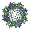
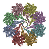
| Title | The cryoEM structure of Monalysin Toxin | |||||||||
 Map data Map data | Reconstruction of Monalysin | |||||||||
 Sample Sample |
| |||||||||
 Keywords Keywords | Monalysin / cryo electron microscopy / single-particle | |||||||||
| Biological species |  | |||||||||
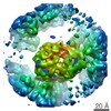
| Method | single particle reconstruction / cryo EM / Resolution: 17.0 Å | |||||||||
 Authors Authors | Leone P / Bebeacua C / Opota O / Kellenberger C / Klaholz B / Cambillau C / Lemaitre B / Roussel A | |||||||||
 Citation Citation | Journal: J Biol Chem / Year: 2015 Title: X-ray and Cryo-electron Microscopy Structures of Monalysin Pore-forming Toxin Reveal Multimerization of the Pro-form. Authors: Philippe Leone / Cecilia Bebeacua / Onya Opota / Christine Kellenberger / Bruno Klaholz / Igor Orlov / Christian Cambillau / Bruno Lemaitre / Alain Roussel /   Abstract: β-Barrel pore-forming toxins (β-PFT), a large family of bacterial toxins, are generally secreted as water-soluble monomers and can form oligomeric pores in membranes following proteolytic cleavage ...β-Barrel pore-forming toxins (β-PFT), a large family of bacterial toxins, are generally secreted as water-soluble monomers and can form oligomeric pores in membranes following proteolytic cleavage and interaction with cell surface receptors. Monalysin has been recently identified as a β-PFT that contributes to the virulence of Pseudomonas entomophila against Drosophila. It is secreted as a pro-protein that becomes active upon cleavage. Here we report the crystal and cryo-electron microscopy structure of the pro-form of Monalysin as well as the crystal structures of the cleaved form and of an inactive mutant lacking the membrane-spanning region. The overall structure of Monalysin displays an elongated shape, which resembles those of β-pore-forming toxins, such as Aerolysin, but is devoid of a receptor-binding domain. X-ray crystallography, cryo-electron microscopy, and light-scattering studies show that pro-Monalysin forms a stable doughnut-like 18-mer complex composed of two disk-shaped nonamers held together by N-terminal swapping of the pro-peptides. This observation is in contrast with the monomeric pro-form of the other β-PFTs that are receptor-dependent for membrane interaction. The membrane-spanning region of pro-Monalysin is fully buried in the center of the doughnut, suggesting that upon cleavage of pro-peptides, the two disk-shaped nonamers can, and have to, dissociate to leave the transmembrane segments free to deploy and lead to pore formation. In contrast with other toxins, the delivery of 18 subunits at once, nearby the cell surface, may be used to bypass the requirement of receptor-dependent concentration to reach the threshold for oligomerization into the pore-forming complex. | |||||||||
| History |
|
- Structure visualization
| Movie |
 Movie viewer Movie viewer |
|---|---|
| Structure viewer | EM map: SurfViewMolmilJmol/JSmol |
| Supplemental images |

- Downloads & links
-EMDB archive
| Map data | emd_2698.map.gz | 8 MB | EMDB map data format | |
|---|---|---|---|---|
| Header (meta data) | emd-2698-v30.xmlemd-2698.xml | 8 KB 8 KB | Display Display | EMDB header |
| Images | 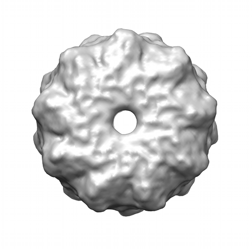 emd_2698.png emd_2698.png | 88.1 KB | ||
| Archive directory |  http://ftp.pdbj.org/pub/emdb/structures/EMD-2698ftp://ftp.pdbj.org/pub/emdb/structures/EMD-2698 http://ftp.pdbj.org/pub/emdb/structures/EMD-2698ftp://ftp.pdbj.org/pub/emdb/structures/EMD-2698 | HTTPS FTP |
-Validation report
| Summary document | emd_2698_validation.pdf.gz | 194.8 KB | Display | EMDB validaton report |
|---|---|---|---|---|
| Full document | emd_2698_full_validation.pdf.gz | 194 KB | Display | |
| Data in XML | emd_2698_validation.xml.gz | 5.8 KB | Display | |
| Arichive directory | https://ftp.pdbj.org/pub/emdb/validation_reports/EMD-2698ftp://ftp.pdbj.org/pub/emdb/validation_reports/EMD-2698 | HTTPS FTP |
-Related structure data


-Links
| EMDB pages | EMDB (EBI/PDBe) / EMDataResource |
|---|
-Map
| File | Download / File: emd_2698.map.gz / Format: CCP4 / Size: 11.1 MB / Type: IMAGE STORED AS FLOATING POINT NUMBER (4 BYTES) | ||||||||||||||||||||||||||||||||||||||||||||||||||||||||||||||||||||
|---|---|---|---|---|---|---|---|---|---|---|---|---|---|---|---|---|---|---|---|---|---|---|---|---|---|---|---|---|---|---|---|---|---|---|---|---|---|---|---|---|---|---|---|---|---|---|---|---|---|---|---|---|---|---|---|---|---|---|---|---|---|---|---|---|---|---|---|---|---|
| Annotation | Reconstruction of Monalysin | ||||||||||||||||||||||||||||||||||||||||||||||||||||||||||||||||||||
| Voxel size | X=Y=Z: 1.92 Å | ||||||||||||||||||||||||||||||||||||||||||||||||||||||||||||||||||||
| Density |
| ||||||||||||||||||||||||||||||||||||||||||||||||||||||||||||||||||||
| Symmetry | Space group: 1 | ||||||||||||||||||||||||||||||||||||||||||||||||||||||||||||||||||||
| Details | EMDB XML:
CCP4 map header:
| ||||||||||||||||||||||||||||||||||||||||||||||||||||||||||||||||||||
-Supplemental data
- Sample components
-Entire : Monalysin
| Entire | Name: Monalysin |
|---|---|
| Components |
|
-Supramolecule #1000: Monalysin
| Supramolecule | Name: Monalysin / type: sample / ID: 1000 / Oligomeric state: 18mer / Number unique components: 1 |
|---|
-Macromolecule #1: Monalysin
| Macromolecule | Name: Monalysin / type: protein_or_peptide / ID: 1 / Number of copies: 9 / Oligomeric state: 9mer / Recombinant expression: Yes |
|---|---|
| Source (natural) | Organism: |
| Recombinant expression | Organism: |
-Experimental details
-Structure determination
| Method | cryo EM |
|---|---|
 Processing Processing | single particle reconstruction |
| Aggregation state | particle |
-Sample preparation
| Concentration | 0.5 mg/mL |
|---|---|
| Buffer | pH: 8 / Details: 10mM Tris, 500mM NaCl |
| Vitrification | Cryogen name: ETHANE / Chamber humidity: 100 % / Instrument: FEI VITROBOT MARK I / Method: Blot for 2 seconds before plunging |
- Electron microscopy
| Microscope | FEI TECNAI F30 |
|---|---|
| Alignment procedure | Legacy - Astigmatism: Objective lens astigmatism was corrected at 100,000 times magnification. |
| Date | Sep 1, 2012 |
| Image recording | Digitization - Sampling interval: 1.92 µm / Number real images: 220 / Average electron dose: 10 e/Å2 |
| Electron beam | Acceleration voltage: 300 kV / Electron source:  FIELD EMISSION GUN FIELD EMISSION GUN |
| Electron optics | Illumination mode: SPOT SCAN / Imaging mode: BRIGHT FIELD / Cs: 2.0 mm / Nominal defocus max: 0.002 µm / Nominal defocus min: 0.001 µm / Nominal magnification: 60000 |
| Sample stage | Specimen holder model: PHILIPS ROTATION HOLDER |
| Experimental equipment |  Model: Tecnai F30 / Image courtesy: FEI Company |
-Image processing
| CTF correction | Details: Images |
|---|---|
| Final reconstruction | Applied symmetry - Point group: D9 (2x9 fold dihedral) / Algorithm: OTHER / Resolution.type: BY AUTHOR / Resolution: 17.0 Å / Resolution method: OTHER / Software - Name: Eman2, Spider, Xmipp / Number images used: 12000 |
