 Movie
Movie Controller

[English] 日本語
 Yorodumi
- EMDB-2658: 20S pre1-1 proteasomal complex from S. cerevisiae carrying the de... -
+ Open data

- Basic information
| Entry | Database: EMDB / ID: EMD-2658 | |||||||||
|---|---|---|---|---|---|---|---|---|---|---|
| Title | 20S pre1-1 proteasomal complex from S. cerevisiae carrying the dedicated assembly chaperones Pba1 and Pba2 | |||||||||
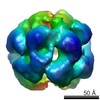
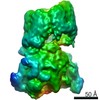
 Map data Map data | 3D Reconstruction of mutant 20S proteasome carrying the dedicated assembly chaperones Pba1 and Pba2 | |||||||||
 Sample Sample |
| |||||||||
 Keywords Keywords | 20S proteasome / Pba1 / Pba2 / pre1-1 | |||||||||
| Biological species |  | |||||||||
| Method | single particle reconstruction / negative staining / Resolution: 21.0 Å | |||||||||
 Authors Authors | Kock M / Nunes MM / Hemann M / Kube S / Dohmen JR / Herzog F / Ramos PC / Wendler P | |||||||||
 Citation Citation | Journal: Nat Commun / Year: 2015 Title: Proteasome assembly from 15S precursors involves major conformational changes and recycling of the Pba1-Pba2 chaperone. Authors: Malte Kock / Maria M Nunes / Matthias Hemann / Sebastian Kube / R Jürgen Dohmen / Franz Herzog / Paula C Ramos / Petra Wendler /  Abstract: The chaperones Ump1 and Pba1-Pba2 promote efficient biogenesis of 20S proteasome core particles from its subunits via 15S intermediates containing alpha and beta subunits, except beta7. Here we ...The chaperones Ump1 and Pba1-Pba2 promote efficient biogenesis of 20S proteasome core particles from its subunits via 15S intermediates containing alpha and beta subunits, except beta7. Here we elucidate the structural role of these chaperones in late steps of core particle biogenesis using biochemical, electron microscopy, cross-linking and mass spectrometry analyses. In 15S precursor complexes, Ump1 is largely unstructured, lining the inner cavity of the complex along the interface between alpha and beta subunits. The alpha and beta subunits form loosely packed rings with a wider alpha ring opening than in the 20S core particle, allowing for the Pba1-Pba2 heterodimer to be partially embedded in the central alpha ring cavity. During biogenesis, the heterodimer is expelled from the alpha ring by a restructuring event that organizes the beta ring and leads to tightening of the alpha ring opening. In this way, the Pba1-Pba2 chaperone is recycled for a new round of proteasome assembly. | |||||||||
| History |
|
- Structure visualization
| Movie |
 Movie viewer Movie viewer |
|---|---|
| Structure viewer | EM map: SurfViewMolmilJmol/JSmol |
| Supplemental images |
 UCSF Chimera
- Downloads & links
-EMDB archive
| Map data | emd_2658.map.gz | 152.9 KB | EMDB map data format | |
|---|---|---|---|---|
| Header (meta data) | emd-2658-v30.xmlemd-2658.xml | 9.4 KB 9.4 KB | Display Display | EMDB header |
| Images | emd_2658.tif | 339.9 KB | ||
| Archive directory |  http://ftp.pdbj.org/pub/emdb/structures/EMD-2658ftp://ftp.pdbj.org/pub/emdb/structures/EMD-2658 http://ftp.pdbj.org/pub/emdb/structures/EMD-2658ftp://ftp.pdbj.org/pub/emdb/structures/EMD-2658 | HTTPS FTP |
-Validation report
| Summary document | emd_2658_validation.pdf.gz | 199.7 KB | Display | EMDB validaton report |
|---|---|---|---|---|
| Full document | emd_2658_full_validation.pdf.gz | 198.9 KB | Display | |
| Data in XML | emd_2658_validation.xml.gz | 5.1 KB | Display | |
| Arichive directory | https://ftp.pdbj.org/pub/emdb/validation_reports/EMD-2658ftp://ftp.pdbj.org/pub/emdb/validation_reports/EMD-2658 | HTTPS FTP |
-Related structure data


-Links
| EMDB pages | EMDB (EBI/PDBe) / EMDataResource |
|---|
-Map
| File | Download / File: emd_2658.map.gz / Format: CCP4 / Size: 3.7 MB / Type: IMAGE STORED AS FLOATING POINT NUMBER (4 BYTES) | ||||||||||||||||||||||||||||||||||||||||||||||||||||||||||||||||||||
|---|---|---|---|---|---|---|---|---|---|---|---|---|---|---|---|---|---|---|---|---|---|---|---|---|---|---|---|---|---|---|---|---|---|---|---|---|---|---|---|---|---|---|---|---|---|---|---|---|---|---|---|---|---|---|---|---|---|---|---|---|---|---|---|---|---|---|---|---|---|
| Annotation | 3D Reconstruction of mutant 20S proteasome carrying the dedicated assembly chaperones Pba1 and Pba2 | ||||||||||||||||||||||||||||||||||||||||||||||||||||||||||||||||||||
| Voxel size | X=Y=Z: 2.9 Å | ||||||||||||||||||||||||||||||||||||||||||||||||||||||||||||||||||||
| Density |
| ||||||||||||||||||||||||||||||||||||||||||||||||||||||||||||||||||||
| Symmetry | Space group: 1 | ||||||||||||||||||||||||||||||||||||||||||||||||||||||||||||||||||||
| Details | EMDB XML:
CCP4 map header:
| ||||||||||||||||||||||||||||||||||||||||||||||||||||||||||||||||||||
-Supplemental data
- Sample components
-Entire : 20S proteasome from S. cerevisiae carrying a pre1-1 mutation as w...
| Entire | Name: 20S proteasome from S. cerevisiae carrying a pre1-1 mutation as well as the dedicated assembly chaperones Pba1 and Pba2 |
|---|---|
| Components |
|
-Supramolecule #1000: 20S proteasome from S. cerevisiae carrying a pre1-1 mutation as w...
| Supramolecule | Name: 20S proteasome from S. cerevisiae carrying a pre1-1 mutation as well as the dedicated assembly chaperones Pba1 and Pba2 type: sample / ID: 1000 / Oligomeric state: Monomer / Number unique components: 1 |
|---|---|
| Molecular weight | Experimental: 830 KDa / Theoretical: 830 KDa |
-Macromolecule #1: 20S pre1-1 proteasomal precursor complex
| Macromolecule | Name: 20S pre1-1 proteasomal precursor complex / type: protein_or_peptide / ID: 1 / Name.synonym: 20S pre1-1 / Number of copies: 1 / Oligomeric state: Monomer / Recombinant expression: No |
|---|---|
| Source (natural) | Organism: |
| Molecular weight | Experimental: 830 KDa / Theoretical: 830 KDa |
-Experimental details
-Structure determination
| Method | negative staining |
|---|---|
 Processing Processing | single particle reconstruction |
| Aggregation state | particle |
-Sample preparation
| Concentration | 0.05 mg/mL |
|---|---|
| Buffer | pH: 7.5 Details: 50 mM Tris-HCl pH 7.5, 5 mM MgCl2, 2 mM ATP, 150 mM NaCl, and 15 % (v/v) glycerol, 10 mM imidazole |
| Staining | Type: NEGATIVE Details: Grids with adsorbed protein floated on 4 drops of 2 % (w/v) uranyl acetate for 10 s each |
| Grid | Details: 400 mesh copper grids with carbon support, glow discharged in ambient atmosphere |
| Vitrification | Cryogen name: NONE / Instrument: OTHER |
- Electron microscopy
| Microscope | FEI TECNAI SPIRIT |
|---|---|
| Alignment procedure | Legacy - Astigmatism: Objective lens astigmatism was corrected at 96000 times magnification |
| Date | Oct 10, 2013 |
| Image recording | Category: CCD / Film or detector model: FEI EAGLE (2k x 2k) / Number real images: 1122 / Average electron dose: 20 e/Å2 |
| Electron beam | Acceleration voltage: 120 kV / Electron source: LAB6 |
| Electron optics | Illumination mode: FLOOD BEAM / Imaging mode: BRIGHT FIELD / Cs: 2.0 mm / Nominal defocus max: 1.0 µm / Nominal defocus min: 0.3 µm / Nominal magnification: 103448 |
| Sample stage | Specimen holder model: SIDE ENTRY, EUCENTRIC |
| Experimental equipment |  Model: Tecnai Spirit / Image courtesy: FEI Company |
-Image processing
| Details | The particles were selected in an automated fashion using Find-EM |
|---|---|
| CTF correction | Details: Micrograph-based |
| Final reconstruction | Applied symmetry - Point group: C1 (asymmetric) / Algorithm: OTHER / Resolution.type: BY AUTHOR / Resolution: 21.0 Å / Resolution method: OTHER / Software - Name: IMAGIC, Spider / Number images used: 12609 |
