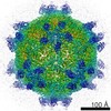
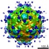
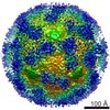
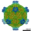
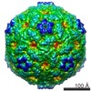
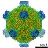
Journal: J Virol / Year: 2014 Title: Cryo-electron microscopy study of insect cell-expressed enterovirus 71 and coxsackievirus a16 virus-like particles provides a structural basis for vaccine development. Authors: Minqing Gong / Hongtao Zhu / Jun Zhou / Chunting Yang / Jing Feng / Xiaojun Huang / Gang Ji / Honglin Xu / Ping Zhu / Abstract: Enterovirus 71 (EV71) and coxsackievirus A16 (CA16) are the two most common etiological agents responsible for the epidemics of hand, foot, and mouth disease (HFMD), a childhood illness with ...Enterovirus 71 (EV71) and coxsackievirus A16 (CA16) are the two most common etiological agents responsible for the epidemics of hand, foot, and mouth disease (HFMD), a childhood illness with occasional severe neurological complications. A number of vaccine candidates against EV71 or CA16 have been reported; however, no vaccine is currently available for clinical use. Here, we generated a secreted version of EV71 and CA16 virus-like particles (VLPs) using a baculovirus-insect cell expression system and reconstructed the three-dimensional (3D) structures of both VLPs by cryo-electron microscopy (cryo-EM) single-particle analysis at 5.2-Å and 5.5-Å resolutions, respectively. The reconstruction results showed that the cryo-EM structures of EV71 and CA16 VLPs highly resemble the recently published crystal structures for EV71 natural empty particles and CA16 135S-like expanded particles, respectively. Our cryo-EM analysis also revealed that the majority of previously identified linear neutralizing epitopes are well preserved on the surface of EV71 and CA16 VLPs. In addition, both VLPs were able to induce efficiently neutralizing antibodies against various strains of EV71 and CA16 viruses in mouse immunization. These studies provide a structural basis for the development of insect cell-expressed VLP vaccines and for a potential bivalent VLP vaccine against both EV71- and CA16-associated HFMD. IMPORTANCE: The recent outbreaks of hand, foot, and mouth disease (HFMD) in the Asia Pacific region spurred the search for effective vaccines against EV71 and CA16 viruses, the two most common ...IMPORTANCE: The recent outbreaks of hand, foot, and mouth disease (HFMD) in the Asia Pacific region spurred the search for effective vaccines against EV71 and CA16 viruses, the two most common etiological agents responsible for HFMD. In this paper, we show that secreted versions of EV71 and CA16 VLPs generated in the baculovirus-insect cell expression system highly resemble the crystal structures of their viral conterparts and that the majority of previously identified linear neutralizing epitopes are well preserved on the VLP surfaces. In addition, the generated VLPs can efficiently induce neutralizing antibodies against various strains of EV71 and CA16 viruses in mouse immunization. These studies provide a structural basis for the development of insect cell-expressed VLP vaccines and for a potential bivalent VLP vaccine against both EV71- and CA16-associated HFMD.
History
Deposition
Mar 12, 2014
-
Header (metadata) release
Apr 16, 2014
-
Map release
Apr 16, 2014
-
Update
May 14, 2014
-
Current status
May 14, 2014
Processing site: PDBe / Status: Released
-
Structure visualization
Movie
Surface view with section colored by density value
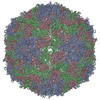
Applied symmetry - Point group: I (icosahedral) / Resolution.type: BY AUTHOR / Resolution: 5.2 Å / Resolution method: OTHER / Number images used: 30386
In the structure databanks used in Yorodumi, some data are registered as the other names, "COVID-19 virus" and "2019-nCoV". Here are the details of the virus and the list of structure data.
Jan 31, 2019. EMDB accession codes are about to change! (news from PDBe EMDB page)
EMDB accession codes are about to change! (news from PDBe EMDB page)
The allocation of 4 digits for EMDB accession codes will soon come to an end. Whilst these codes will remain in use, new EMDB accession codes will include an additional digit and will expand incrementally as the available range of codes is exhausted. The current 4-digit format prefixed with “EMD-” (i.e. EMD-XXXX) will advance to a 5-digit format (i.e. EMD-XXXXX), and so on. It is currently estimated that the 4-digit codes will be depleted around Spring 2019, at which point the 5-digit format will come into force.
The EM Navigator/Yorodumi systems omit the EMD- prefix.
Related info.:Q: What is EMD? / ID/Accession-code notation in Yorodumi/EM Navigator
Yorodumi is a browser for structure data from EMDB, PDB, SASBDB, etc.
This page is also the successor to EM Navigator detail page, and also detail information page/front-end page for Omokage search.
The word "yorodu" (or yorozu) is an old Japanese word meaning "ten thousand". "mi" (miru) is to see.
Related info.:EMDB / PDB / SASBDB / Comparison of 3 databanks / Yorodumi Search / Aug 31, 2016. New EM Navigator & Yorodumi / Yorodumi Papers / Jmol/JSmol / Function and homology information / Changes in new EM Navigator and Yorodumi
 Movie
Movie Controller

 Yorodumi
 Open data
 Basic information
 Map data
 Sample
 Human enterovirus 71
 Authors
 Citation
 Structure visualization
 Movie viewer
 Downloads & links
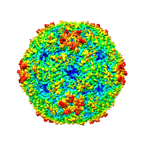 emd_2607.png
 http://ftp.pdbj.org/pub/emdb/structures/EMD-2607
 Sample components
 Homo sapiens (human) / synonym: VERTEBRATES
 Spodoptera frugiperda (fall armyworm) / Recombinant cell: Sf9 / Recombinant plasmid: pFastBac Dual
 Processing
 Electron microscopy
 FIELD EMISSION GUN