 Movie
Movie Controller

[English] 日本語
 Yorodumi
- EMDB-2490: Structure of the 39S Large Subunit of the Mammalian Mitochondrial... -
+ Open data

- Basic information
| Entry | Database: EMDB / ID: EMD-2490 | |||||||||
|---|---|---|---|---|---|---|---|---|---|---|
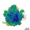
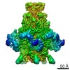
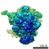
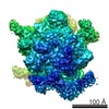
| Title | Structure of the 39S Large Subunit of the Mammalian Mitochondrial Ribosome | |||||||||
 Map data Map data | Structure of the 39S Large Subunit of the Mammalian Mitochondrial Ribosome | |||||||||
 Sample Sample |
| |||||||||
 Keywords Keywords | mammalian mitochondrial ribosome / 39S large ribosomal subunit /  translation / ribosomal proteins / rRNA / polypeptide exit site / membrane association translation / ribosomal proteins / rRNA / polypeptide exit site / membrane association | |||||||||
| Function / homology |  Function and homology information Function and homology informationMitochondrial translation elongation / Mitochondrial translation termination / translation release factor activity / rRNA import into mitochondrion / mitochondrial translational elongation / microprocessor complex / ribonuclease III activity / mitochondrial large ribosomal subunit / peptidyl-tRNA hydrolase / mitochondrial translation ...Mitochondrial translation elongation / Mitochondrial translation termination / translation release factor activity / rRNA import into mitochondrion / mitochondrial translational elongation / microprocessor complex / ribonuclease III activity / mitochondrial large ribosomal subunit / peptidyl-tRNA hydrolase / mitochondrial translation / organelle membrane / RNA processing / double-stranded RNA binding / large ribosomal subunit / 5S rRNA binding / rRNA binding / nuclear body / ribosome / structural constituent of ribosome / ribonucleoprotein complex / translation / protein domain specific binding / nucleotide binding / intracellular membrane-bounded organelle / mitochondrion / RNA binding / nucleus / plasma membrane / cytosol / cytoplasmSimilarity search - Function | |||||||||
| Biological species |  Sus scrofa domesticus (domestic pig) Sus scrofa domesticus (domestic pig) | |||||||||
| Method | single particle reconstruction / cryo EM / Resolution: 4.9 Å | |||||||||
 Authors Authors | Greber BJ / Boehringer D / Leitner A / Bieri P / Voigts-Hoffmann F / Erzberger JP / Leibundgut M / Aebersold R / Ban N | |||||||||
 Citation Citation | Journal: Nature / Year: 2014 Title: Architecture of the large subunit of the mammalian mitochondrial ribosome. Authors: Basil J Greber / Daniel Boehringer / Alexander Leitner / Philipp Bieri / Felix Voigts-Hoffmann / Jan P Erzberger / Marc Leibundgut / Ruedi Aebersold / Nenad Ban /  Abstract: Mitochondrial ribosomes synthesize a number of highly hydrophobic proteins encoded on the genome of mitochondria, the organelles in eukaryotic cells that are responsible for energy conversion by ...Mitochondrial ribosomes synthesize a number of highly hydrophobic proteins encoded on the genome of mitochondria, the organelles in eukaryotic cells that are responsible for energy conversion by oxidative phosphorylation. The ribosomes in mammalian mitochondria have undergone massive structural changes throughout their evolution, including ribosomal RNA shortening and acquisition of mitochondria-specific ribosomal proteins. Here we present the three-dimensional structure of the 39S large subunit of the porcine mitochondrial ribosome determined by cryo-electron microscopy at 4.9 Å resolution. The structure, combined with data from chemical crosslinking and mass spectrometry experiments, reveals the unique features of the 39S subunit at near-atomic resolution and provides detailed insight into the architecture of the polypeptide exit site. This region of the mitochondrial ribosome has been considerably remodelled compared to its bacterial counterpart, providing a specialized platform for the synthesis and membrane insertion of the highly hydrophobic protein components of the respiratory chain. | |||||||||
| History |
|
- Structure visualization
| Movie |
Movie viewer |
|---|---|
| Structure viewer | EM map: SurfViewMolmilJmol/JSmol |
| Supplemental images |

- Downloads & links
-EMDB archive
| Map data | emd_2490.map.gz | 59.9 MB | EMDB map data format | |
|---|---|---|---|---|
| Header (meta data) | emd-2490-v30.xmlemd-2490.xml | 20.8 KB 20.8 KB | Display Display | EMDB header |
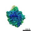
| Images | 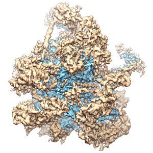 EMD-2490-image_EMDB_ray_500px.png EMD-2490-image_EMDB_ray_500px.png | 129.6 KB | ||
| Archive directory |  http://ftp.pdbj.org/pub/emdb/structures/EMD-2490ftp://ftp.pdbj.org/pub/emdb/structures/EMD-2490 http://ftp.pdbj.org/pub/emdb/structures/EMD-2490ftp://ftp.pdbj.org/pub/emdb/structures/EMD-2490 | HTTPS FTP |
-Related structure data
| Related structure data |  4ce4MC M: atomic model generated by this map C: citing same article ( |
|---|---|
| Similar structure data |
-Links
| EMDB pages | EMDB (EBI/PDBe) / EMDataResource |
|---|---|
| Related items in Molecule of the Month |


-Map
| File | Download / File: emd_2490.map.gz / Format: CCP4 / Size: 62.5 MB / Type: IMAGE STORED AS FLOATING POINT NUMBER (4 BYTES) | ||||||||||||||||||||||||||||||||||||||||||||||||||||||||||||||||||||
|---|---|---|---|---|---|---|---|---|---|---|---|---|---|---|---|---|---|---|---|---|---|---|---|---|---|---|---|---|---|---|---|---|---|---|---|---|---|---|---|---|---|---|---|---|---|---|---|---|---|---|---|---|---|---|---|---|---|---|---|---|---|---|---|---|---|---|---|---|---|
| Annotation | Structure of the 39S Large Subunit of the Mammalian Mitochondrial Ribosome | ||||||||||||||||||||||||||||||||||||||||||||||||||||||||||||||||||||
| Voxel size | X=Y=Z: 1.41 Å | ||||||||||||||||||||||||||||||||||||||||||||||||||||||||||||||||||||
| Density |
| ||||||||||||||||||||||||||||||||||||||||||||||||||||||||||||||||||||
| Symmetry | Space group: 1 | ||||||||||||||||||||||||||||||||||||||||||||||||||||||||||||||||||||
| Details | EMDB XML:
CCP4 map header:
| ||||||||||||||||||||||||||||||||||||||||||||||||||||||||||||||||||||
-Supplemental data
- Sample components
-Entire : 39S large subunit of the porcine mitochondrial ribosome
| Entire | Name: 39S large subunit of the porcine mitochondrial ribosome |
|---|---|
| Components |
|
-Supramolecule #1000: 39S large subunit of the porcine mitochondrial ribosome
| Supramolecule | Name: 39S large subunit of the porcine mitochondrial ribosome type: sample / ID: 1000 / Oligomeric state: monomer / Number unique components: 1 |
|---|---|
| Molecular weight | Theoretical: 1.6 MDa |
-Supramolecule #1: 39S large subunit of the mitochondrial ribosome
| Supramolecule | Name: 39S large subunit of the mitochondrial ribosome / type: organelle_or_cellular_component / ID: 1 / Number of copies: 1 / Oligomeric state: Monomer / Recombinant expression: No |
|---|---|
| Ref GO | divclassse qspanoncli ckpopupspa nclassgree n(this)spandata popltspanc lassquotlo adingbarqu otgtltimgs rcquotimgl oadinggifq uotdecodin gquotasync quotgtltsp angtdataur lajaxphp?m odetaxoamp ... divclassse qspanoncli ckpopupspa nclassgree n(this)spandata popltspanc lassquotlo adingbarqu otgtltimgs rcquotimgl oadinggifq uotdecodin gquotasync quotgtltsp angtdataur lajaxphp?m odetaxoamp kGO3A00057 61ampajax1 classpoptr giGO000576 1ispandiv |
| Source (natural) | Organism: Sus scrofa domesticus (domestic pig) / synonym: domestic pig / Tissue: Liver / Organelle: Mitochondrion |
| Molecular weight | Theoretical: 1.6 MDa |
 Processing
 Electron microscopy #1

