 Movie
Movie Controller

[English] 日本語
 Yorodumi
- EMDB-2374: cryo-electron microscopy of Sindbis-liposome complex at pH 6.4 (t... -
+ Open data

- Basic information
| Entry | Database: EMDB / ID: EMD-2374 | |||||||||
|---|---|---|---|---|---|---|---|---|---|---|
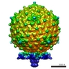
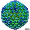
| Title | cryo-electron microscopy of Sindbis-liposome complex at pH 6.4 (the group of 5-fold reconstruction) | |||||||||
 Map data Map data | reconstruction of Sindbis virus when a liposome associates to its 50-fold axis. 5-fold symmetry was applied to the reconstruction map. | |||||||||
 Sample Sample |
| |||||||||
 Keywords Keywords |  cryo-electron microscopy / 3D reconstruction / Sindbis virus / membrane fusion / low pH cryo-electron microscopy / 3D reconstruction / Sindbis virus / membrane fusion / low pH | |||||||||
| Biological species |  Sindbis virus Sindbis virus | |||||||||
| Method | single particle reconstruction / cryo EM / Resolution: 14.5 Å | |||||||||
 Authors Authors | Cao S / Zhang W | |||||||||
 Citation Citation | Journal: Proc Natl Acad Sci U S A / Year: 2013 Title: Characterization of an early-stage fusion intermediate of Sindbis virus using cryoelectron microscopy. Authors: Sheng Cao / Wei Zhang /  Abstract: The sequential steps in the alphavirus membrane fusion pathway have been postulated based on the prefusion and postfusion crystal structures of the viral fusion protein E1 in conjunction with ...The sequential steps in the alphavirus membrane fusion pathway have been postulated based on the prefusion and postfusion crystal structures of the viral fusion protein E1 in conjunction with biochemical studies. However, the molecular structures of the hypothesized fusion intermediates have remained obscure due to difficulties inherent in the dynamic nature of the process. We developed an experimental system that uses liposomes as the target membrane to capture Sindbis virus, a prototypical alphavirus, in its membrane-binding form at pH 6.4. Cryoelectron micrograph analyses and 3D reconstructions showed that the virus retains its overall icosahedral structure at this mildly acidic pH, except in the membrane-binding region, where monomeric E1 associates with the target membrane and the E2 glycoprotein retains its original trimeric organization. The remaining E2 trimers may hinder E1 homotrimerization and are a potential target for antiviral drugs. | |||||||||
| History |
|
- Structure visualization
| Movie |
 Movie viewer Movie viewer |
|---|---|
| Structure viewer | EM map: SurfViewMolmilJmol/JSmol |
| Supplemental images |

- Downloads & links
-EMDB archive
| Map data | emd_2374.map.gz | 25.3 MB | EMDB map data format | |
|---|---|---|---|---|
| Header (meta data) | emd-2374-v30.xmlemd-2374.xml | 8.5 KB 8.5 KB | Display Display | EMDB header |
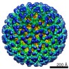
| Images | 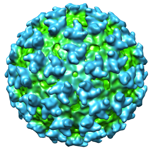 image2374.png image2374.png | 287.8 KB | ||
| Archive directory |  http://ftp.pdbj.org/pub/emdb/structures/EMD-2374ftp://ftp.pdbj.org/pub/emdb/structures/EMD-2374 http://ftp.pdbj.org/pub/emdb/structures/EMD-2374ftp://ftp.pdbj.org/pub/emdb/structures/EMD-2374 | HTTPS FTP |
-Related structure data
| Similar structure data |
|---|
-Links
| EMDB pages | EMDB (EBI/PDBe) / EMDataResource |
|---|
-Map
| File | Download / File: emd_2374.map.gz / Format: CCP4 / Size: 61.8 MB / Type: IMAGE STORED AS FLOATING POINT NUMBER (4 BYTES) | ||||||||||||||||||||||||||||||||||||||||||||||||||||||||||||||||||||
|---|---|---|---|---|---|---|---|---|---|---|---|---|---|---|---|---|---|---|---|---|---|---|---|---|---|---|---|---|---|---|---|---|---|---|---|---|---|---|---|---|---|---|---|---|---|---|---|---|---|---|---|---|---|---|---|---|---|---|---|---|---|---|---|---|---|---|---|---|---|
| Annotation | reconstruction of Sindbis virus when a liposome associates to its 50-fold axis. 5-fold symmetry was applied to the reconstruction map. | ||||||||||||||||||||||||||||||||||||||||||||||||||||||||||||||||||||
| Voxel size | X=Y=Z: 3.951 Å | ||||||||||||||||||||||||||||||||||||||||||||||||||||||||||||||||||||
| Density |
| ||||||||||||||||||||||||||||||||||||||||||||||||||||||||||||||||||||
| Symmetry | Space group: 1 | ||||||||||||||||||||||||||||||||||||||||||||||||||||||||||||||||||||
| Details | EMDB XML:
CCP4 map header:
| ||||||||||||||||||||||||||||||||||||||||||||||||||||||||||||||||||||
-Supplemental data
- Sample components
-Entire : Sindbis virus - liposome complex at pH 6.4
| Entire | Name: Sindbis virus - liposome complex at pH 6.4 |
|---|---|
| Components |
|
-Supramolecule #1000: Sindbis virus - liposome complex at pH 6.4
| Supramolecule | Name: Sindbis virus - liposome complex at pH 6.4 / type: sample / ID: 1000 / Number unique components: 1 |
|---|
-Supramolecule #1: Sindbis virus
| Supramolecule | Name: Sindbis virus / type: virus / ID: 1 Details: The virus was mixed with liposome and was acidified to pH 6.4 at 37C for 2 min NCBI-ID: 11034 / Sci species name: Sindbis virus / Sci species strain: TE12 / Virus type: VIRION / Virus isolate: STRAIN / Virus enveloped: Yes / Virus empty: No |
|---|---|
| Host (natural) | Organism:  Homo sapiens (human) / synonym: VERTEBRATES Homo sapiens (human) / synonym: VERTEBRATES |
| Host system | Organism:  Mesocricetus auratus (golden hamster) / Recombinant cell: BHK-21 Mesocricetus auratus (golden hamster) / Recombinant cell: BHK-21 |
| Virus shell | Shell ID: 1 / Name: glycoprotein / Diameter: 700 Å / T number (triangulation number): 4 |
-Experimental details
-Structure determination
| Method | cryo EM |
|---|---|
 Processing Processing | single particle reconstruction |
| Aggregation state | particle |
-Sample preparation
| Buffer | pH: 6.4 Details: Virus and liposome were suspended in TNE (10mM Tris, 200mM NaCl, 1mM EDTA, pH 7.4). The low pH buffer is (50mM MES, 200mM NaCl, pH.6.4) |
|---|---|
| Grid | Details: Ultra-thin carbon film |
| Vitrification | Cryogen name: ETHANE / Chamber humidity: 100 % / Instrument: FEI VITROBOT MARK III Timed resolved state: Vitrified 2 min after waiting in the chamber (37C) |
- Electron microscopy
| Microscope | FEI TECNAI F30 |
|---|---|
| Electron beam | Acceleration voltage: 300 kV / Electron source: FIELD EMISSION GUN |
| Electron optics | Calibrated magnification: 75930 / Illumination mode: FLOOD BEAM / Imaging mode: BRIGHT FIELDBright-field microscopy / Cs: 2.0 mm / Nominal defocus max: 4.0 µm / Nominal defocus min: 1.0 µm / Nominal magnification: 59000 |
| Sample stage | Specimen holder model: GATAN LIQUID NITROGEN / Tilt angle min: 0 / Tilt angle max: 0 |
| Temperature | Average: 95 K |
| Date | Feb 4, 2012 |
| Image recording | Category: CCD / Film or detector model: GATAN ULTRASCAN 4000 (4k x 4k) / Average electron dose: 25 e/Å2 |
| Experimental equipment |  Model: Tecnai F30 / Image courtesy: FEI Company |
-Image processing
| CTF correction | Details: each particle |
|---|---|
| Final reconstruction | Applied symmetry - Point group: C5 (5 fold cyclic) / Algorithm: OTHER / Resolution.type: BY AUTHOR / Resolution: 14.5 Å / Resolution method: FSC 0.5 CUT-OFF / Software - Name: auto3dem / Number images used: 196 |
