 Movie
Movie Controller


+ Open data

- Basic information
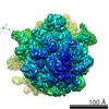
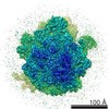
| Entry | Database: EMDB / ID: EMD-2373 | |||||||||
|---|---|---|---|---|---|---|---|---|---|---|
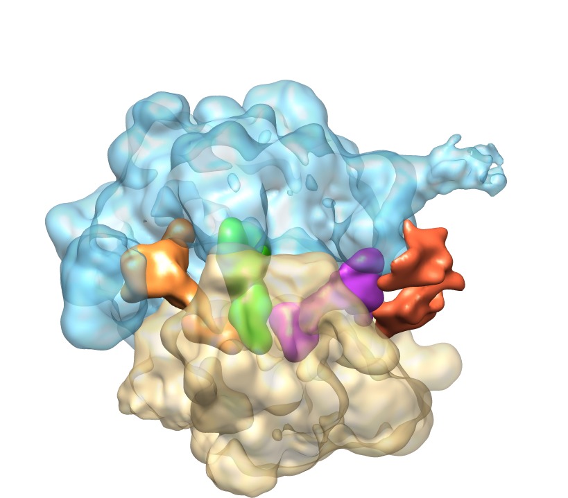
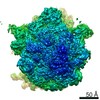
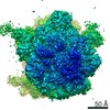
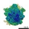
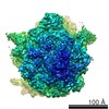
| Title | ribosome-RelA complex | |||||||||
 Map data Map data | reconstruction of ribosome-RelA complex | |||||||||
 Sample Sample |
| |||||||||
 Keywords Keywords |  ribosome / RelA / stringent response ribosome / RelA / stringent response | |||||||||
| Biological species |   Thermus thermophilus (bacteria) / Escherichia coli (E. coli) Thermus thermophilus (bacteria) / Escherichia coli (E. coli) | |||||||||
| Method | single particle reconstruction / cryo EM / negative staining / Resolution: 14.5 Å | |||||||||
 Authors Authors | Agirrezabala X / Fernandez I / Kelley A / Gil-Carton D / Ramakrishnan V / Valle M | |||||||||
 Citation Citation | Journal: EMBO Rep / Year: 2013 Title: The ribosome triggers the stringent response by RelA via a highly distorted tRNA. Authors: Xabier Agirrezabala / Israel S Fernández / Ann C Kelley / David Gil Cartón / Venki Ramakrishnan / Mikel Valle /  Abstract: The bacterial stringent response links nutrient starvation with the transcriptional control of genes. This process is initiated by the stringent factor RelA, which senses the presence of deacylated ...The bacterial stringent response links nutrient starvation with the transcriptional control of genes. This process is initiated by the stringent factor RelA, which senses the presence of deacylated tRNA in the ribosome as a symptom of amino-acid starvation to synthesize the alarmone (p)ppGpp. Here we report a cryo-EM study of RelA bound to ribosomes bearing cognate, deacylated tRNA in the A-site. The data show that RelA on the ribosome stabilizes an unusual distorted form of the tRNA, with the acceptor arm making contact with RelA and far from its normal location in the peptidyl transferase centre. | |||||||||
| History |
|
- Structure visualization
| Movie |
 Movie viewer Movie viewer |
|---|---|
| Structure viewer | EM map: SurfViewMolmilJmol/JSmol |
| Supplemental images |
- Downloads & links
-EMDB archive
| Map data | emd_2373.map.gz | 33 MB | EMDB map data format | |
|---|---|---|---|---|
| Header (meta data) | emd-2373-v30.xmlemd-2373.xml | 11.6 KB 11.6 KB | Display Display | EMDB header |
| Images | EMD-2373-4.tif | 455.1 KB | ||
| Archive directory |  http://ftp.pdbj.org/pub/emdb/structures/EMD-2373ftp://ftp.pdbj.org/pub/emdb/structures/EMD-2373 http://ftp.pdbj.org/pub/emdb/structures/EMD-2373ftp://ftp.pdbj.org/pub/emdb/structures/EMD-2373 | HTTPS FTP |
-Related structure data
| Similar structure data |
|---|

-Links
| EMDB pages | EMDB (EBI/PDBe) / EMDataResource |
|---|---|
| Related items in Molecule of the Month |

-Map
| File | Download / File: emd_2373.map.gz / Format: CCP4 / Size: 34.5 MB / Type: IMAGE STORED AS FLOATING POINT NUMBER (4 BYTES) | ||||||||||||||||||||||||||||||||||||||||||||||||||||||||||||||||||||
|---|---|---|---|---|---|---|---|---|---|---|---|---|---|---|---|---|---|---|---|---|---|---|---|---|---|---|---|---|---|---|---|---|---|---|---|---|---|---|---|---|---|---|---|---|---|---|---|---|---|---|---|---|---|---|---|---|---|---|---|---|---|---|---|---|---|---|---|---|---|
| Annotation | reconstruction of ribosome-RelA complex | ||||||||||||||||||||||||||||||||||||||||||||||||||||||||||||||||||||
| Voxel size | X=Y=Z: 1.75 Å | ||||||||||||||||||||||||||||||||||||||||||||||||||||||||||||||||||||
| Density |
| ||||||||||||||||||||||||||||||||||||||||||||||||||||||||||||||||||||
| Symmetry | Space group: 1 | ||||||||||||||||||||||||||||||||||||||||||||||||||||||||||||||||||||
| Details | EMDB XML:
CCP4 map header:
| ||||||||||||||||||||||||||||||||||||||||||||||||||||||||||||||||||||
-Supplemental data
- Sample components
-Entire : 70S-RelA complex
| Entire | Name: 70S-RelA complex |
|---|---|
| Components |
|
-Supramolecule #1000: 70S-RelA complex
| Supramolecule | Name: 70S-RelA complex / type: sample / ID: 1000 / Number unique components: 5 |
|---|---|
| Molecular weight | Theoretical: 2.5 MDa |
-Supramolecule #1: 70S ribosome
| Supramolecule | Name: 70S ribosome / type: complex / ID: 1 / Recombinant expression: No / Ribosome-details: ribosome-prokaryote: ALL |
|---|---|
| Source (natural) | Organism: Thermus thermophilus (bacteria) |
| Molecular weight | Theoretical: 2.5 MDa |
-Macromolecule #1: deacylated tRNA
| Macromolecule | Name: deacylated tRNA / type: rna / ID: 1 / Classification: OTHER / Structure: SINGLE STRANDED / Synthetic?: No |
|---|---|
| Source (natural) | Organism: Thermus thermophilus (bacteria) |
-Macromolecule #2: tRNA
| Macromolecule | Name: tRNA / type: rna / ID: 2 / Classification: OTHER / Structure: SINGLE STRANDED / Synthetic?: No |
|---|---|
| Source (natural) | Organism: Thermus thermophilus (bacteria) |
-Macromolecule #3: tRNA
| Macromolecule | Name: tRNA / type: rna / ID: 3 / Classification: OTHER / Structure: SINGLE STRANDED / Synthetic?: No |
|---|---|
| Source (natural) | Organism: Thermus thermophilus (bacteria) |
-Macromolecule #4: RelA
| Macromolecule | Name: RelA / type: protein_or_peptide / ID: 4 / Recombinant expression: No |
|---|---|
| Source (natural) | Organism: Escherichia coli (E. coli) |
-Experimental details
-Structure determination
| Method | negative staining, cryo EM |
|---|---|
 Processing Processing | single particle reconstruction |
| Aggregation state | particle |
-Sample preparation
| Concentration | 0.25 mg/mL |
|---|---|
| Buffer | pH: 7.45 Details: 5mM Hepes-KOH pH 7.45, 50mM KCl, 10mM NH4Cl, 10mM Mg-acetate, 5mM beta-mercapto-ethanol |
| Staining | Type: NEGATIVE / Details: cryo |
| Grid | Details: Quantifoil grids (2/4) with thin carbon on top |
| Vitrification | Cryogen name: ETHANE / Chamber humidity: 100 % / Chamber temperature: 90 K / Instrument: FEI VITROBOT MARK I / Method: Blot for 3 seconds before plunging |
- Electron microscopy
| Microscope | JEOL 2200FS |
|---|---|
| Electron beam | Acceleration voltage: 200 kV / Electron source: FIELD EMISSION GUN |
| Electron optics | Calibrated magnification: 40000 / Illumination mode: FLOOD BEAM / Imaging mode: BRIGHT FIELDBright-field microscopy / Cs: 2 mm / Nominal defocus max: 4.0 µm / Nominal defocus min: 1.0 µm / Nominal magnification: 40000 |
| Specialist optics | Energy filter - Name: in-column Omega filter / Energy filter - Lower energy threshold: 15.0 eV / Energy filter - Upper energy threshold: 20.0 eV |
| Sample stage | Specimen holder model: GATAN LIQUID NITROGEN |
| Alignment procedure | Legacy - Astigmatism: corrected at 100k |
| Date | Jan 15, 2012 |
| Image recording | Category: FILM / Film or detector model: KODAK SO-163 FILM / Digitization - Scanner: OTHER / Digitization - Sampling interval: 7 µm / Number real images: 425 / Average electron dose: 10 e/Å2 / Bits/pixel: 8 |
-Image processing
| CTF correction | Details: defocus groups |
|---|---|
| Final reconstruction | Applied symmetry - Point group: C1 (asymmetric) / Algorithm: OTHER / Resolution.type: BY AUTHOR / Resolution: 14.5 Å / Resolution method: FSC 0.5 CUT-OFF / Software - Name: XMIPP, SPIDER / Number images used: 28791 |
| Details | particles were selected by combining automated particle picking , multivariate data analysis and visual inspection |
-Atomic model buiding 1
| Initial model | PDB ID:  2wrn |
|---|---|
| Software | Name: MDFF |
| Refinement | Space: REAL / Protocol: FLEXIBLE FIT |
-Atomic model buiding 2
| Initial model | PDB ID: 2wro |
|---|---|
| Software | Name: MDFF |
| Refinement | Space: REAL / Protocol: FLEXIBLE FIT |
