Movie
Movie Controller


+ Open data

- Basic information
| Entry | Database: EMDB / ID: EMD-2347 | |||||||||
|---|---|---|---|---|---|---|---|---|---|---|
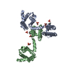
| Title | cryo-EM structure of the NavCt voltage-gated sodium channel | |||||||||
 Map data Map data | Cryo-EM structure of voltage-gated Na+ channel | |||||||||
 Sample Sample |
| |||||||||
 Keywords Keywords | Voltage-gated sodium ion channel / tetrameric ion channel | |||||||||
| Function / homology | Voltage-dependent channel domain superfamily /  Ion transport domain / Ion transport domain / Ion transport protein / monoatomic ion channel activity / membrane / Ion transport protein Ion transport domain / Ion transport domain / Ion transport protein / monoatomic ion channel activity / membrane / Ion transport protein Function and homology information Function and homology information | |||||||||
| Biological species |   Caldalkalibacillus thermarum (bacteria) Caldalkalibacillus thermarum (bacteria) | |||||||||
| Method | electron crystallography / cryo EM / Resolution: 9.0 Å | |||||||||
 Authors Authors | Tsai C-J / Tani K / Irie K / Hiroaki Y / Shimomura T / McMillan DG / Cook GM / Schertler G / Fujiyoshi Y / Li X-D | |||||||||
 Citation Citation | Journal: J Mol Biol / Year: 2013 Title: Two alternative conformations of a voltage-gated sodium channel. Authors: Ching-Ju Tsai / Kazutoshi Tani / Katsumasa Irie / Yoko Hiroaki / Takushi Shimomura / Duncan G McMillan / Gregory M Cook / Gebhard F X Schertler / Yoshinori Fujiyoshi / Xiao-Dan Li /  Abstract: Activation and inactivation of voltage-gated sodium channels (Navs) are well studied, yet the molecular mechanisms governing channel gating in the membrane remain unknown. We present two ...Activation and inactivation of voltage-gated sodium channels (Navs) are well studied, yet the molecular mechanisms governing channel gating in the membrane remain unknown. We present two conformations of a Nav from Caldalkalibacillus thermarum reconstituted into lipid bilayers in one crystal at 9Å resolution based on electron crystallography. Despite a voltage sensor arrangement identical with that in the activated form, we observed two distinct pore domain structures: a prominent form with a relatively open inner gate and a closed inner-gate conformation similar to the first prokaryotic Nav structure. Structural differences, together with mutational and electrophysiological analyses, indicated that widening of the inner gate was dependent on interactions among the S4-S5 linker, the N-terminal part of S5 and its adjoining part in S6, and on interhelical repulsion by a negatively charged C-terminal region subsequent to S6. Our findings suggest that these specific interactions result in two conformational structures. | |||||||||
| History |
|
- Structure visualization
| Movie |
Movie viewer |
|---|---|
| Structure viewer | EM map: SurfViewMolmilJmol/JSmol |
| Supplemental images |

- Downloads & links
-EMDB archive
| Map data | emd_2347.map.gz | 545.4 KB | EMDB map data format | |
|---|---|---|---|---|
| Header (meta data) | emd-2347-v30.xmlemd-2347.xml | 11.7 KB 11.7 KB | Display Display | EMDB header |
| Images |  EMD-2347.jpg EMD-2347.jpg | 177.6 KB | ||
| Archive directory |  http://ftp.pdbj.org/pub/emdb/structures/EMD-2347ftp://ftp.pdbj.org/pub/emdb/structures/EMD-2347 http://ftp.pdbj.org/pub/emdb/structures/EMD-2347ftp://ftp.pdbj.org/pub/emdb/structures/EMD-2347 | HTTPS FTP |
-Related structure data
| Related structure data |  4bgnMC M: atomic model generated by this map C: citing same article ( |
|---|---|
| Similar structure data |
-Links
| EMDB pages | EMDB (EBI/PDBe) / EMDataResource |
|---|---|
| Related items in Molecule of the Month |

-Map
| File | Download / File: emd_2347.map.gz / Format: CCP4 / Size: 936.5 KB / Type: IMAGE STORED AS FLOATING POINT NUMBER (4 BYTES) | ||||||||||||||||||||||||||||||||||||||||||||||||||||||||||||||||||||
|---|---|---|---|---|---|---|---|---|---|---|---|---|---|---|---|---|---|---|---|---|---|---|---|---|---|---|---|---|---|---|---|---|---|---|---|---|---|---|---|---|---|---|---|---|---|---|---|---|---|---|---|---|---|---|---|---|---|---|---|---|---|---|---|---|---|---|---|---|---|
| Annotation | Cryo-EM structure of voltage-gated Na+ channel | ||||||||||||||||||||||||||||||||||||||||||||||||||||||||||||||||||||
| Voxel size | X: 2.09091 Å / Y: 2.09091 Å / Z: 2.22222 Å | ||||||||||||||||||||||||||||||||||||||||||||||||||||||||||||||||||||
| Density |
| ||||||||||||||||||||||||||||||||||||||||||||||||||||||||||||||||||||
| Symmetry | Space group: 1 | ||||||||||||||||||||||||||||||||||||||||||||||||||||||||||||||||||||
| Details | EMDB XML:
CCP4 map header:
| ||||||||||||||||||||||||||||||||||||||||||||||||||||||||||||||||||||
-Supplemental data
- Sample components
-Entire : voltage-gated sodium channel
| Entire | Name: voltage-gated sodium channelSodium channel |
|---|---|
| Components |
|
-Supramolecule #1000: voltage-gated sodium channel
| Supramolecule | Name: voltage-gated sodium channel / type: sample / ID: 1000 Oligomeric state: Two tetramers of voltage-gated sodium channel Number unique components: 1 |
|---|
-Macromolecule #1: voltage-gated sodium channel
| Macromolecule | Name: voltage-gated sodium channel / type: protein_or_peptide / ID: 1 / Number of copies: 2 / Oligomeric state: tetramer / Recombinant expression: Yes |
|---|---|
| Source (natural) | Organism: Caldalkalibacillus thermarum (bacteria) / Strain: TA2.A1 / Location in cell: Plasma membrane |
| Recombinant expression | Organism: Escherichia coli BL21(DE3) (bacteria) / Recombinant strain: C43 / Recombinant plasmid: pTrc99A |
| Sequence | UniProtKB: Ion transport protein / GO: monoatomic ion channel activity / InterPro: Ion transport domain |
-Experimental details
-Structure determination
| Method | cryo EM |
|---|---|
 Processing Processing | electron crystallography |
| Aggregation state | 2D array |
-Sample preparation
| Concentration | 4.0 mg/mL |
|---|---|
| Buffer | pH: 9 Details: 50mM glycine-NaOH pH9.0, 200mM NaCl, 4mM MgCl2, 5% glycerol, 5% methyl-2,4-pentanediol, 1.5mM NaN3 |
| Grid | Details: molybdenum EM grid |
| Vitrification | Cryogen name: NITROGEN / Instrument: LEICA KF80 |
| Details | Crystals grown by dialysis |
| Crystal formation | Details: Crystals grown by dialysis |
- Electron microscopy
| Microscope | JEOL KYOTO-3000SFF |
|---|---|
| Electron beam | Acceleration voltage: 300 kV / Electron source: FIELD EMISSION GUN |
| Electron optics | Calibrated magnification: 39500 / Illumination mode: FLOOD BEAM / Imaging mode: BRIGHT FIELDBright-field microscopy / Cs: 1.6 mm / Nominal defocus max: 3.82 µm / Nominal defocus min: 0.91 µm / Nominal magnification: 40000 |
| Sample stage | Specimen holder: Helium cooled, top entry / Specimen holder model: JEOL / Tilt angle max: 60 / Tilt series - Axis1 - Min angle: 0 ° / Tilt series - Axis1 - Max angle: 60 ° |
| Temperature | Min: 4 K / Average: 4 K |
| Date | Nov 5, 2010 |
| Image recording | Category: FILM / Film or detector model: KODAK SO-163 FILM / Digitization - Scanner: ZEISS SCAI / Digitization - Sampling interval: 7 µm / Number real images: 77 / Average electron dose: 20 e/Å2 / Bits/pixel: 14 |
| Tilt angle min | 0 |
-Image processing
| Crystal parameters | Unit cell - A: 115.0 Å / Unit cell - B: 115.0 Å / Unit cell - C: 180.0 Å / Unit cell - γ: 90.0 ° / Unit cell - α: 90.0 ° / Unit cell - β: 90.0 ° / Plane group: P 4 |
|---|---|
| CTF correction | Details: Each micrographs |
| Final reconstruction | Resolution.type: BY AUTHOR / Resolution: 9.0 Å / Resolution method: OTHER / Software - Name: MRC |
| Details | Images were processed using MRC suite |