 Movie
Movie Controller

[English] 日本語
 Yorodumi
- EMDB-2340: The electron microscopy reconstruction of the baseplate of lactoc... -
+ Open data

- Basic information
| Entry | Database: EMDB / ID: EMD-2340 | |||||||||
|---|---|---|---|---|---|---|---|---|---|---|
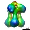
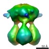
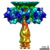
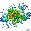
| Title | The electron microscopy reconstruction of the baseplate of lactococcal phage Tuc2009. | |||||||||
 Map data Map data | Reconstruction of the baseplate of lactococcal phage Tuc2009 | |||||||||
 Sample Sample |
| |||||||||
 Keywords Keywords | lactococcal phage / tuc2009 /  baseplate / electron microscopy / single-particle baseplate / electron microscopy / single-particle | |||||||||
| Biological species |  Lactococcus phage Tuc2009 (virus) Lactococcus phage Tuc2009 (virus) | |||||||||
| Method | single particle reconstruction / negative staining / Resolution: 36.0 Å | |||||||||
 Authors Authors | Collins B / Bebeacua C / Mahony J / Douillard F / Veesler D / Blangy B / Cambillau C / van Sinderen D | |||||||||
 Citation Citation | Journal: J Virol / Year: 2013 Title: Structure and functional analysis of the host recognition device of lactococcal phage tuc2009. Authors: Barry Collins / Cecilia Bebeacua / Jennifer Mahony / Stéphanie Blangy / François P Douillard / David Veesler / Christian Cambillau / Douwe van Sinderen /  Abstract: Many phages employ a large heteropolymeric organelle located at the tip of the tail, termed the baseplate, for host recognition. Contrast electron microscopy (EM) of the lactococcal phage Tuc2009 ...Many phages employ a large heteropolymeric organelle located at the tip of the tail, termed the baseplate, for host recognition. Contrast electron microscopy (EM) of the lactococcal phage Tuc2009 baseplate and its host-binding subunits, the so-called tripods, allowed us to obtain a low-resolution structural image of this organelle. Structural comparisons between the baseplate of the related phage TP901-1 and that of Tuc2009 demonstrated that they are highly similar, except for the presence of an additional protein in the Tuc2009 baseplate (BppATuc2009), which is attached to the top of the Tuc2009 tripod structure. Recombinantly produced Tuc2009 or TP901-1 tripods were shown to bind specifically to their particular host cell surfaces and are capable of almost fully and specifically eliminating Tuc2009 or TP901-1 phage adsorption, respectively. In the case of Tuc2009, such adsorption-blocking ability was reduced in tripods that lacked BppATuc2009, indicating that this protein increases the binding specificity and/or affinity of the Tuc2009 tripod to its host receptor. | |||||||||
| History |
|
- Structure visualization
| Movie |
 Movie viewer Movie viewer |
|---|---|
| Structure viewer | EM map: SurfViewMolmilJmol/JSmol |
| Supplemental images |

- Downloads & links
-EMDB archive
| Map data | emd_2340.map.gz | 8.8 MB | EMDB map data format | |
|---|---|---|---|---|
| Header (meta data) | emd-2340-v30.xmlemd-2340.xml | 9.4 KB 9.4 KB | Display Display | EMDB header |
| Images | 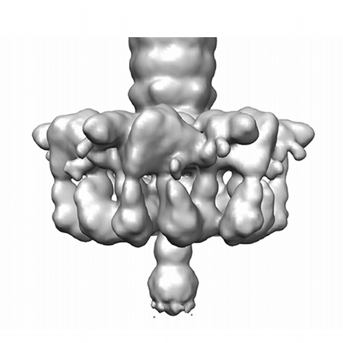 emd_2340.png emd_2340.png | 103.3 KB | ||
| Archive directory |  http://ftp.pdbj.org/pub/emdb/structures/EMD-2340ftp://ftp.pdbj.org/pub/emdb/structures/EMD-2340 http://ftp.pdbj.org/pub/emdb/structures/EMD-2340ftp://ftp.pdbj.org/pub/emdb/structures/EMD-2340 | HTTPS FTP |
-Related structure data


-Links
| EMDB pages | EMDB (EBI/PDBe) / EMDataResource |
|---|
-Map
| File | Download / File: emd_2340.map.gz / Format: CCP4 / Size: 11.1 MB / Type: IMAGE STORED AS FLOATING POINT NUMBER (4 BYTES) | ||||||||||||||||||||||||||||||||||||||||||||||||||||||||||||||||||||
|---|---|---|---|---|---|---|---|---|---|---|---|---|---|---|---|---|---|---|---|---|---|---|---|---|---|---|---|---|---|---|---|---|---|---|---|---|---|---|---|---|---|---|---|---|---|---|---|---|---|---|---|---|---|---|---|---|---|---|---|---|---|---|---|---|---|---|---|---|---|
| Annotation | Reconstruction of the baseplate of lactococcal phage Tuc2009 | ||||||||||||||||||||||||||||||||||||||||||||||||||||||||||||||||||||
| Voxel size | X=Y=Z: 4.95 Å | ||||||||||||||||||||||||||||||||||||||||||||||||||||||||||||||||||||
| Density |
| ||||||||||||||||||||||||||||||||||||||||||||||||||||||||||||||||||||
| Symmetry | Space group: 1 | ||||||||||||||||||||||||||||||||||||||||||||||||||||||||||||||||||||
| Details | EMDB XML:
CCP4 map header:
| ||||||||||||||||||||||||||||||||||||||||||||||||||||||||||||||||||||
-Supplemental data
- Sample components
-Entire : Baseplate of lactoccocal phage Tuc2009
| Entire | Name: Baseplate of lactoccocal phage Tuc2009 |
|---|---|
| Components |
|
-Supramolecule #1000: Baseplate of lactoccocal phage Tuc2009
| Supramolecule | Name: Baseplate of lactoccocal phage Tuc2009 / type: sample / ID: 1000 Details: The baseplate was selected from images collected from a sample of the entire phages. Oligomeric state: 6 / Number unique components: 1 |
|---|
-Supramolecule #1: Lactococcus phage Tuc2009
| Supramolecule | Name: Lactococcus phage Tuc2009 / type: virus / ID: 1 / Name.synonym: Tuc2009 / NCBI-ID: 35241 / Sci species name: Lactococcus phage Tuc2009 / Virus type: OTHER / Virus isolate: SPECIES / Virus enveloped: No / Virus empty: No / Syn species name: Tuc2009 |
|---|---|
| Host (natural) | Organism:  Lactococcus lactis (lactic acid bacteria) / Strain: UC509.9 / synonym: BACTERIA(EUBACTERIA) Lactococcus lactis (lactic acid bacteria) / Strain: UC509.9 / synonym: BACTERIA(EUBACTERIA) |
-Experimental details
-Structure determination
| Method | negative staining |
|---|---|
 Processing Processing | single particle reconstruction |
| Aggregation state | particle |
-Sample preparation
| Buffer | pH: 7.4 / Details: 20 mM Tris-HCL, 10 mM MgSO4, 100 mM NaCl |
|---|---|
| Staining | Type: NEGATIVE Details: Grids with adsorbed protein floated on 2% w/v uranyl acetate for 20 seconds |
| Grid | Details: 300 mesh copper grid with thin carbon layer, glow discharged for 30 seconds |
| Vitrification | Cryogen name: NONE / Instrument: OTHER |
- Electron microscopy
| Microscope | FEI TECNAI 12 |
|---|---|
| Electron beam | Acceleration voltage: 120 kV / Electron source: LAB6 |
| Electron optics | Calibrated magnification: 48500 / Illumination mode: FLOOD BEAM / Imaging mode: BRIGHT FIELDBright-field microscopy / Cs: 2 mm / Nominal defocus max: 1.5 µm / Nominal defocus min: 1.5 µm / Nominal magnification: 48500 |
| Sample stage | Specimen holder: Room temperature holder / Specimen holder model: SIDE ENTRY, EUCENTRIC |
| Alignment procedure | Legacy - Astigmatism: Objective lens astigmatism was corrected at 100,000 times magnification |
| Date | Jun 1, 2012 |
| Image recording | Category: CCD / Film or detector model: GENERIC CCD / Number real images: 2000 / Average electron dose: 10 e/Å2 / Details: Images were collected using a 2kx2k CCD camera |
-Image processing
| Final reconstruction | Applied symmetry - Point group: C6 (6 fold cyclic) / Algorithm: OTHER / Resolution.type: BY AUTHOR / Resolution: 36.0 Å / Resolution method: OTHER / Software - Name: EMAN2, Spider, Xmipp / Number images used: 2000 |
|---|
-Atomic model buiding 1
| Initial model | PDB ID:  4div |
|---|---|
| Software | Name: Chimera |
| Details | The baseplate was manually fitted and then automatically refined |
| Refinement | Space: REAL / Protocol: RIGID BODY FIT |
