Movie
Movie Controller

[English] 日本語
 Yorodumi
- EMDB-2327: Visualizing GroEL/ES in the Act of Encapsulating a Non-Native Sub... -
+ Open data

- Basic information
| Entry | Database: EMDB / ID: EMD-2327 | |||||||||
|---|---|---|---|---|---|---|---|---|---|---|
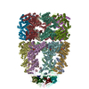
| Title | Visualizing GroEL/ES in the Act of Encapsulating a Non-Native Substrate Protein | |||||||||
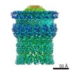
 Map data Map data | Symmetry-free 3D reconstruction of the bullet-shaped substrate-encapsulated subpopulation sorted from the heterogeneous sample of wild-type GroEL reacted with the substrate protein RuBisCO, GroES and nucleotide ATP | |||||||||
 Sample Sample |
| |||||||||
 Keywords Keywords |  protein folding / chaperonin / GroEL / GroEL-GroES / cryo-EM / heterogeneity / substrate / RuBisCO / encapsulation protein folding / chaperonin / GroEL / GroEL-GroES / cryo-EM / heterogeneity / substrate / RuBisCO / encapsulation | |||||||||
| Function / homology |  Function and homology information Function and homology informationGroEL-GroES complex / chaperonin ATPase / virion assembly / chaperone cofactor-dependent protein refolding / protein folding chaperone / isomerase activity / ATP-dependent protein folding chaperone / response to radiation / unfolded protein binding / protein folding ...GroEL-GroES complex / chaperonin ATPase / virion assembly / chaperone cofactor-dependent protein refolding / protein folding chaperone / isomerase activity / ATP-dependent protein folding chaperone / response to radiation / unfolded protein binding / protein folding / response to heat / protein-folding chaperone binding / protein refolding / magnesium ion binding / ATP hydrolysis activity / ATP binding / membrane / identical protein binding / metal ion binding / cytosolSimilarity search - Function | |||||||||
| Biological species |  Escherichia coli K-12 (bacteria) / Rhodospirillum rubrum (bacteria) / synthetic construct (others) Escherichia coli K-12 (bacteria) / Rhodospirillum rubrum (bacteria) / synthetic construct (others) | |||||||||
| Method | single particle reconstruction / cryo EM / Resolution: 15.9 Å | |||||||||
 Authors Authors | Chen D-H / Madan D / Weaver J / Lin Z / Schroder GF / Chiu W / Rye HS | |||||||||
 Citation Citation | Journal: Cell / Year: 2013 Title: Visualizing GroEL/ES in the act of encapsulating a folding protein. Authors: Dong-Hua Chen / Damian Madan / Jeremy Weaver / Zong Lin / Gunnar F Schröder / Wah Chiu / Hays S Rye /  Abstract: The GroEL/ES chaperonin system is required for the assisted folding of many proteins. How these substrate proteins are encapsulated within the GroEL-GroES cavity is poorly understood. Using symmetry- ...The GroEL/ES chaperonin system is required for the assisted folding of many proteins. How these substrate proteins are encapsulated within the GroEL-GroES cavity is poorly understood. Using symmetry-free, single-particle cryo-electron microscopy, we have characterized a chemically modified mutant of GroEL (EL43Py) that is trapped at a normally transient stage of substrate protein encapsulation. We show that the symmetric pattern of the GroEL subunits is broken as the GroEL cis-ring apical domains reorient to accommodate the simultaneous binding of GroES and an incompletely folded substrate protein (RuBisCO). The collapsed RuBisCO folding intermediate binds to the lower segment of two apical domains, as well as to the normally unstructured GroEL C-terminal tails. A comparative structural analysis suggests that the allosteric transitions leading to substrate protein release and folding involve concerted shifts of GroES and the GroEL apical domains and C-terminal tails. | |||||||||
| History |
|
- Structure visualization
| Movie |
Movie viewer |
|---|---|
| Structure viewer | EM map: SurfViewMolmilJmol/JSmol |
| Supplemental images |

- Downloads & links
-EMDB archive
| Map data | emd_2327.map.gz | 19.3 MB | EMDB map data format | |
|---|---|---|---|---|
| Header (meta data) | emd-2327-v30.xmlemd-2327.xml | 16.6 KB 16.6 KB | Display Display | EMDB header |
| Images | 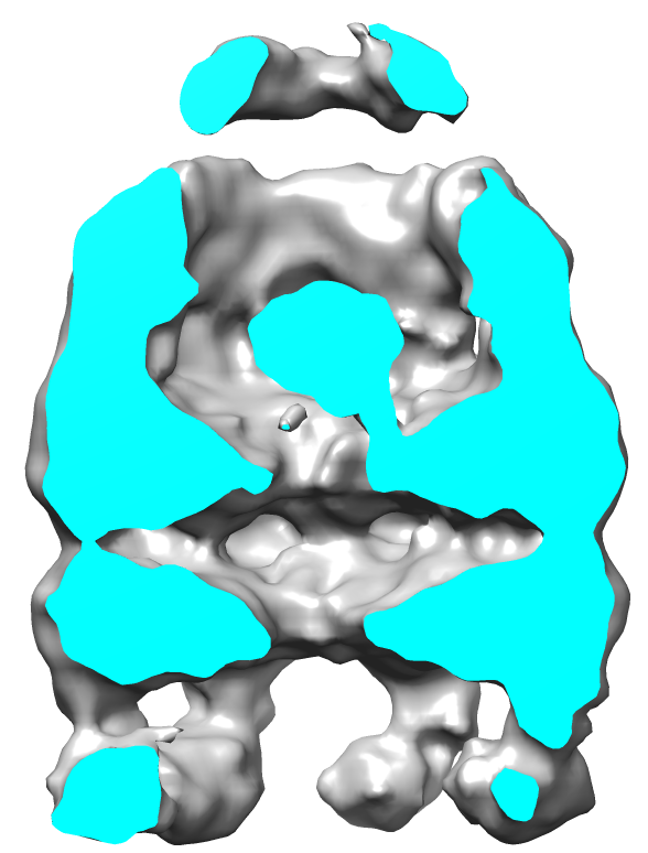 EMD-2327.png EMD-2327.png | 135.8 KB | ||
| Archive directory |  http://ftp.pdbj.org/pub/emdb/structures/EMD-2327ftp://ftp.pdbj.org/pub/emdb/structures/EMD-2327 http://ftp.pdbj.org/pub/emdb/structures/EMD-2327ftp://ftp.pdbj.org/pub/emdb/structures/EMD-2327 | HTTPS FTP |
-Related structure data
| Related structure data | 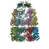 3zq1MC 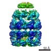 2325C 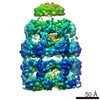 2326C 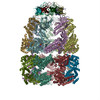 3zpzC  3zq0C M: atomic model generated by this map C: citing same article ( |
|---|---|
| Similar structure data |


-Links
| EMDB pages | EMDB (EBI/PDBe) / EMDataResource |
|---|---|
| Related items in Molecule of the Month |


-Map
| File | Download / File: emd_2327.map.gz / Format: CCP4 / Size: 21.7 MB / Type: IMAGE STORED AS FLOATING POINT NUMBER (4 BYTES) | ||||||||||||||||||||||||||||||||||||||||||||||||||||||||||||||||||||
|---|---|---|---|---|---|---|---|---|---|---|---|---|---|---|---|---|---|---|---|---|---|---|---|---|---|---|---|---|---|---|---|---|---|---|---|---|---|---|---|---|---|---|---|---|---|---|---|---|---|---|---|---|---|---|---|---|---|---|---|---|---|---|---|---|---|---|---|---|---|
| Annotation | Symmetry-free 3D reconstruction of the bullet-shaped substrate-encapsulated subpopulation sorted from the heterogeneous sample of wild-type GroEL reacted with the substrate protein RuBisCO, GroES and nucleotide ATP | ||||||||||||||||||||||||||||||||||||||||||||||||||||||||||||||||||||
| Voxel size | X=Y=Z: 2.12 Å | ||||||||||||||||||||||||||||||||||||||||||||||||||||||||||||||||||||
| Density |
| ||||||||||||||||||||||||||||||||||||||||||||||||||||||||||||||||||||
| Symmetry | Space group: 1 | ||||||||||||||||||||||||||||||||||||||||||||||||||||||||||||||||||||
| Details | EMDB XML:
CCP4 map header:
| ||||||||||||||||||||||||||||||||||||||||||||||||||||||||||||||||||||
-Supplemental data
- Sample components
-Entire : Non-native RuBisCO substrate protein encapsulated inside the cavi...
| Entire | Name: Non-native RuBisCO substrate protein encapsulated inside the cavity of GroEL capped by GroES with the assistance of nucleotide ATP |
|---|---|
| Components |
|
-Supramolecule #1000: Non-native RuBisCO substrate protein encapsulated inside the cavi...
| Supramolecule | Name: Non-native RuBisCO substrate protein encapsulated inside the cavity of GroEL capped by GroES with the assistance of nucleotide ATP type: sample / ID: 1000 / Details: The molecule is bullet-shaped Oligomeric state: Non-native RuBisCO substrate protein was encapsulated inside the cavity formed by one tetradecamer of GroEL, one heptamer of GroES and seven nucleotides of ATP Number unique components: 4 |
|---|---|
| Molecular weight | Experimental: 920 KDa / Theoretical: 920 KDa Method: Estimated by the sum of GroEL molecular weight 800kDa, GroES molecular weight 70kDa and one RuBisCO monomer molecular weight 50kDa |
-Macromolecule #1: GroEL-D398A
| Macromolecule | Name: GroEL-D398A / type: protein_or_peptide / ID: 1 / Name.synonym: EL398A Details: The D398A mutation prevents ATP hydrolysis by GroEL. Number of copies: 14 / Oligomeric state: Tetradecamer / Recombinant expression: Yes |
|---|---|
| Source (natural) | Organism: Escherichia coli K-12 (bacteria) / Strain: BL21 / Location in cell: Cytoplasm |
| Molecular weight | Experimental: 800 KDa / Theoretical: 800 KDa |
| Recombinant expression | Organism: Escherichia coli BL21(DE3) (bacteria) / Recombinant plasmid: pACYC |
| Sequence | UniProtKB: Chaperonin GroEL |
-Macromolecule #2: GroES
| Macromolecule | Name: GroES / type: protein_or_peptide / ID: 2 / Number of copies: 7 / Oligomeric state: Heptamer / Recombinant expression: Yes |
|---|---|
| Source (natural) | Organism: Escherichia coli K-12 (bacteria) / Location in cell: Cytoplasm |
| Molecular weight | Experimental: 70 KDa / Theoretical: 70 KDa |
| Recombinant expression | Organism: Escherichia coli BL21(DE3) (bacteria) / Recombinant plasmid: pACYC |
| Sequence | UniProtKB: Co-chaperonin GroES |
-Macromolecule #3: Ribulose-1,5-bisphosphate carboxylase oxygenase
| Macromolecule | Name: Ribulose-1,5-bisphosphate carboxylase oxygenase / type: protein_or_peptide / ID: 3 / Name.synonym: RuBisCO Details: The native RuBisCO is a dimer, but the encapsulated RuBisCO is a non-native monomer. Number of copies: 1 / Oligomeric state: monomer / Recombinant expression: Yes |
|---|---|
| Source (natural) | Organism: Rhodospirillum rubrum (bacteria) / Location in cell: cytoplasm |
| Molecular weight | Experimental: 50 KDa / Theoretical: 50 KDa |
| Recombinant expression | Organism: Escherichia coli BL21(DE3) (bacteria) / Recombinant plasmid: pUC derived plasmid with T7 promoter |
-Macromolecule #4: Adenosine triphosphate
| Macromolecule | Name: Adenosine triphosphate / type: ligand / ID: 4 / Name.synonym: ATP / Recombinant expression: No |
|---|---|
| Source (natural) | Organism: synthetic construct (others) |
-Experimental details
-Structure determination
| Method | cryo EM |
|---|---|
 Processing Processing | single particle reconstruction |
| Aggregation state | particle |
-Sample preparation
| Concentration | 2.4 mg/mL |
|---|---|
| Buffer | pH: 7.6 / Details: 50 mM Hepes, 50 mM KOAc, 10 mM Mg(OAc)2, 2 mM DTT |
| Grid | Details: 400 mesh R1.2/1.3 Quantifoil grid glow-discharged 10 sec before freezing |
| Vitrification | Cryogen name: ETHANE / Chamber humidity: 95 % / Chamber temperature: 98 K / Instrument: FEI VITROBOT MARK III / Method: Blot for 1 second before plunging |
- Electron microscopy
| Microscope | JEOL 3200FSC |
|---|---|
| Electron beam | Acceleration voltage: 300 kV / Electron source: FIELD EMISSION GUN |
| Electron optics | Calibrated magnification: 70760 / Illumination mode: FLOOD BEAM / Imaging mode: OTHER / Nominal defocus max: 5.0 µm / Nominal defocus min: 1.5 µm / Nominal magnification: 50000 |
| Specialist optics | Energy filter - Name: JEOL / Energy filter - Lower energy threshold: 0.0 eV / Energy filter - Upper energy threshold: 25.0 eV |
| Sample stage | Specimen holder model: JEOL 3200FSC CRYOHOLDER |
| Temperature | Min: 100.9 K / Max: 101.1 K / Average: 101 K |
| Alignment procedure | Legacy - Astigmatism: Objective lens astigmatism was corrected at 100,000 times magnification |
| Date | Dec 21, 2008 |
| Image recording | Category: CCD / Film or detector model: GENERIC GATAN / Digitization - Sampling interval: 15 µm / Number real images: 1537 / Average electron dose: 20 e/Å2 / Bits/pixel: 8 |
-Image processing
| CTF correction | Details: Each frame |
|---|---|
| Final two d classification | Number classes: 280 |
| Final angle assignment | Details: EMAN1 |
| Final reconstruction | Applied symmetry - Point group: C1 (asymmetric) / Algorithm: OTHER / Resolution.type: BY AUTHOR / Resolution: 15.9 Å / Resolution method: OTHER / Software - Name: EMAN1 / Number images used: 8189 |
| Details | Particle selection was performed semi-automatically using the EMAN1 program, boxer. The contrast transfer function fitting was performed automatically using the program fitctf.py and then fine-tuned manually using the EMAN1 program ctfit. The methodology of EMAN1 multiple-model refinement (EMAN1 program multirefine) for compositionally and conformationally heterogeneous complex analysis was used to sort out the relatively homogeneous bullet-shaped particle images with the substrate protein inside the cis cavity. |
-Atomic model buiding 1
| Initial model | PDB ID: Chain - #0 - Chain ID: A / Chain - #1 - Chain ID: B / Chain - #2 - Chain ID: C / Chain - #3 - Chain ID: D / Chain - #4 - Chain ID: E / Chain - #5 - Chain ID: F / Chain - #6 - Chain ID: G / Chain - #7 - Chain ID: H / Chain - #8 - Chain ID: I / Chain - #9 - Chain ID: J / Chain - #10 - Chain ID: K / Chain - #11 - Chain ID: L / Chain - #12 - Chain ID: M / Chain - #13 - Chain ID: N / Chain - #14 - Chain ID: O / Chain - #15 - Chain ID: P / Chain - #16 - Chain ID: Q / Chain - #17 - Chain ID: R / Chain - #18 - Chain ID: S / Chain - #19 - Chain ID: T / Chain - #20 - Chain ID: U |
|---|---|
| Software | Name: DireX |
| Refinement | Space: REAL / Protocol: FLEXIBLE FIT / Target criteria: cross-correlation coefficient |
| Output model | PDB-3zq1: |