Movie
Movie Controller

[English] 日本語
 Yorodumi
- EMDB-2318: Single particle cryo-microscopy reconstruction of the isolated Ma... -
+ Open data

- Basic information
| Entry | Database: EMDB / ID: EMD-2318 | |||||||||
|---|---|---|---|---|---|---|---|---|---|---|
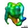
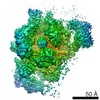
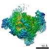
| Title | Single particle cryo-microscopy reconstruction of the isolated Manduca sexta V1 domain | |||||||||
 Map data Map data | V1 domain without the C subunit bound | |||||||||
 Sample Sample |
| |||||||||
 Keywords Keywords |  V-ATPase / atp synthase / membrane complex V-ATPase / atp synthase / membrane complex | |||||||||
| Biological species |  Manduca sexta (tobacco hornworm) Manduca sexta (tobacco hornworm) | |||||||||
| Method | single particle reconstruction / cryo EM / negative staining / Resolution: 20.0 Å | |||||||||
 Authors Authors | Muench SP / Scheres SHW / Huss M / Phillips C / Vitavska O / Wieczorek H / Trinick J / Harrison MA | |||||||||
 Citation Citation | Journal: J Mol Biol / Year: 2014 Title: Subunit positioning and stator filament stiffness in regulation and power transmission in the V1 motor of the Manduca sexta V-ATPase. Authors: Stephen P Muench / Sjors H W Scheres / Markus Huss / Clair Phillips / Olga Vitavska / Helmut Wieczorek / John Trinick / Michael A Harrison /   Abstract: The vacuolar H(+)-ATPase (V-ATPase) is an ATP-driven proton pump essential to the function of eukaryotic cells. Its cytoplasmic V1 domain is an ATPase, normally coupled to membrane-bound proton pump ...The vacuolar H(+)-ATPase (V-ATPase) is an ATP-driven proton pump essential to the function of eukaryotic cells. Its cytoplasmic V1 domain is an ATPase, normally coupled to membrane-bound proton pump Vo via a rotary mechanism. How these asymmetric motors are coupled remains poorly understood. Low energy status can trigger release of V1 from the membrane and curtail ATP hydrolysis. To investigate the molecular basis for these processes, we have carried out cryo-electron microscopy three-dimensional reconstruction of deactivated V1 from Manduca sexta. In the resulting model, three peripheral stalks that are parts of the mechanical stator of the V-ATPase are clearly resolved as unsupported filaments in the same conformations as in the holoenzyme. They are likely therefore to have inherent stiffness consistent with a role as flexible rods in buffering elastic power transmission between the domains of the V-ATPase. Inactivated V1 adopted a homogeneous resting state with one open active site adjacent to the stator filament normally linked to the H subunit. Although present at 1:1 stoichiometry with V1, both recombinant subunit C reconstituted with V1 and its endogenous subunit H were poorly resolved in three-dimensional reconstructions, suggesting structural heterogeneity in the region at the base of V1 that could indicate positional variability. If the position of H can vary, existing mechanistic models of deactivation in which it binds to and locks the axle of the V-ATPase rotary motor would need to be re-evaluated. | |||||||||
| History |
|
- Structure visualization
| Movie |
 Movie viewer Movie viewer |
|---|---|
| Structure viewer | EM map: SurfViewMolmilJmol/JSmol |
| Supplemental images |

- Downloads & links
-EMDB archive
| Map data | emd_2318.map.gz | 461.2 KB | EMDB map data format | |
|---|---|---|---|---|
| Header (meta data) | emd-2318-v30.xmlemd-2318.xml | 11.6 KB 11.6 KB | Display Display | EMDB header |
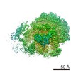
| Images |  image2318.png image2318.png | 112.6 KB | ||
| Archive directory |  http://ftp.pdbj.org/pub/emdb/structures/EMD-2318ftp://ftp.pdbj.org/pub/emdb/structures/EMD-2318 http://ftp.pdbj.org/pub/emdb/structures/EMD-2318ftp://ftp.pdbj.org/pub/emdb/structures/EMD-2318 | HTTPS FTP |
-Related structure data


-Links
| EMDB pages | EMDB (EBI/PDBe) / EMDataResource |
|---|
-Map
| File | Download / File: emd_2318.map.gz / Format: CCP4 / Size: 825.2 KB / Type: IMAGE STORED AS FLOATING POINT NUMBER (4 BYTES) | ||||||||||||||||||||||||||||||||||||||||||||||||||||||||||||||||||||
|---|---|---|---|---|---|---|---|---|---|---|---|---|---|---|---|---|---|---|---|---|---|---|---|---|---|---|---|---|---|---|---|---|---|---|---|---|---|---|---|---|---|---|---|---|---|---|---|---|---|---|---|---|---|---|---|---|---|---|---|---|---|---|---|---|---|---|---|---|---|
| Annotation | V1 domain without the C subunit bound | ||||||||||||||||||||||||||||||||||||||||||||||||||||||||||||||||||||
| Voxel size | X=Y=Z: 4.36 Å | ||||||||||||||||||||||||||||||||||||||||||||||||||||||||||||||||||||
| Density |
| ||||||||||||||||||||||||||||||||||||||||||||||||||||||||||||||||||||
| Symmetry | Space group: 1 | ||||||||||||||||||||||||||||||||||||||||||||||||||||||||||||||||||||
| Details | EMDB XML:
CCP4 map header:
| ||||||||||||||||||||||||||||||||||||||||||||||||||||||||||||||||||||
-Supplemental data
- Sample components
-Entire : M. sexta isolated V1 domain without subunit C
| Entire | Name: M. sexta isolated V1 domain without subunit C |
|---|---|
| Components |
|
-Supramolecule #1000: M. sexta isolated V1 domain without subunit C
| Supramolecule | Name: M. sexta isolated V1 domain without subunit C / type: sample / ID: 1000 Details: The sample was monodisperse and not freeze thawed to maintain V1 integrity Oligomeric state: One V1 bound to a monomer of subunit C / Number unique components: 1 |
|---|---|
| Molecular weight | Experimental: 650 KDa / Theoretical: 650 KDa / Method: Mass Spec |
-Macromolecule #1: Vacuolar ATPase isolated V1 domain
| Macromolecule | Name: Vacuolar ATPase isolated V1 domain / type: protein_or_peptide / ID: 1 / Name.synonym: V1 / Details: No C subunit / Number of copies: 1 / Oligomeric state: 3A,B,E,G subunits and 1 D,F,H subunit / Recombinant expression: No |
|---|---|
| Source (natural) | Organism: Manduca sexta (tobacco hornworm) / synonym: Tobacco Hornworm / Tissue: midgut epithelium |
| Molecular weight | Experimental: 650 KDa / Theoretical: 650 KDa |
-Experimental details
-Structure determination
| Method | negative staining, cryo EM |
|---|---|
 Processing Processing | single particle reconstruction |
| Aggregation state | particle |
-Sample preparation
| Concentration | 1 mg/mL |
|---|---|
| Buffer | pH: 8.1 / Details: 150mM NaCl, 20mM Tris-HCL, 0.01% C12E10 |
| Staining | Type: NEGATIVE Details: negative stain data was collected using 1% w/v uranyl acetate for 1 minute. |
| Grid | Details: UV glow discharged grids, 400 mesh |
| Vitrification | Cryogen name: ETHANE / Chamber humidity: 100 % / Chamber temperature: 120 K / Instrument: FEI VITROBOT MARK IV Method: Grids were blotted for 6 seconds with a 6 second drain time |
- Electron microscopy
| Microscope | FEI TECNAI F20 |
|---|---|
| Electron beam | Acceleration voltage: 200 kV / Electron source: FIELD EMISSION GUN |
| Electron optics | Calibrated magnification: 69000 / Illumination mode: FLOOD BEAM / Imaging mode: BRIGHT FIELDBright-field microscopy / Cs: 2.0 mm / Nominal defocus max: 0.0035 µm / Nominal defocus min: 0.001 µm / Nominal magnification: 50000 |
| Sample stage | Specimen holder: Nitrogen cooled 3350 holder / Specimen holder model: GATAN LIQUID NITROGEN |
| Temperature | Min: 90 K / Max: 110 K / Average: 100 K |
| Alignment procedure | Legacy - Astigmatism: Astigmatism corrected for each image in focus mode at 100,000 times magnification |
| Details | FEI Low dose mode |
| Date | Apr 14, 2011 |
| Image recording | Category: CCD / Film or detector model: GENERIC GATAN (4k x 4k) / Average electron dose: 16 e/Å2 / Details: All collected on Gatan 4K x 4K CCD |
| Experimental equipment |  Model: Tecnai F20 / Image courtesy: FEI Company |
-Image processing
| CTF correction | Details: Relion |
|---|---|
| Final angle assignment | Details: Relion angular sampling 10 degrees |
| Final reconstruction | Applied symmetry - Point group: C1 (asymmetric) / Algorithm: OTHER / Resolution.type: BY AUTHOR / Resolution: 20.0 Å / Resolution method: OTHER / Software - Name: Relion Details: Maximum likelihood in Relion using the MLF3D protocol Number images used: 16500 |
| Details | The particles were handpicked in BOXER |
-Atomic model buiding 1
| Initial model | PDB ID: Chain - #0 - Chain ID: A / Chain - #1 - Chain ID: B / Chain - #2 - Chain ID: C / Chain - #3 - Chain ID: D / Chain - #4 - Chain ID: E / Chain - #5 - Chain ID: F / Chain - #6 - Chain ID: G / Chain - #7 - Chain ID: H |
|---|---|
| Software | Name: Chimera |
| Details | The domains were fitted using Chimera |
| Refinement | Space: REAL / Protocol: RIGID BODY FIT / Target criteria: best fit |