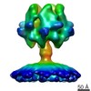
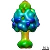
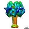
Journal: Nature / Year: 2013 Title: A syringe-like injection mechanism in Photorhabdus luminescens toxins. Authors: Christos Gatsogiannis / Alexander E Lang / Dominic Meusch / Vanda Pfaumann / Oliver Hofnagel / Roland Benz / Klaus Aktories / Stefan Raunser / Abstract: Photorhabdus luminescens is an insect pathogenic bacterium that is symbiotic with entomopathogenic nematodes. On invasion of insect larvae, P. luminescens is released from the nematodes and kills ...Photorhabdus luminescens is an insect pathogenic bacterium that is symbiotic with entomopathogenic nematodes. On invasion of insect larvae, P. luminescens is released from the nematodes and kills the insect through the action of a variety of virulence factors including large tripartite ABC-type toxin complexes (Tcs). Tcs are typically composed of TcA, TcB and TcC proteins and are biologically active only when complete. Functioning as ADP-ribosyltransferases, TcC proteins were identified as the actual functional components that induce actin-clustering, defects in phagocytosis and cell death. However, little is known about the translocation of TcC into the cell by the TcA and TcB components. Here we show that TcA in P. luminescens (TcdA1) forms a transmembrane pore and report its structure in the prepore and pore state determined by cryoelectron microscopy. We find that the TcdA1 prepore assembles as a pentamer forming an α-helical, vuvuzela-shaped channel less than 1.5 nanometres in diameter surrounded by a large outer shell. Membrane insertion is triggered not only at low pH as expected, but also at high pH, explaining Tc action directly through the midgut of insects. Comparisons with structures of the TcdA1 pore inserted into a membrane and in complex with TcdB2 and TccC3 reveal large conformational changes during membrane insertion, suggesting a novel syringe-like mechanism of protein translocation. Our results demonstrate how ABC-type toxin complexes bridge a membrane to insert their lethal components into the cytoplasm of the host cell. We believe that the proposed mechanism is characteristic of the whole ABC-type toxin family. This explanation of toxin translocation is a step towards understanding the host-pathogen interaction and the complex life cycle of P. luminescens and other pathogens, including human pathogenic bacteria, and serves as a strong foundation for the development of biopesticides.
History
Deposition
Jan 25, 2013
-
Header (metadata) release
Feb 13, 2013
-
Map release
Feb 13, 2013
-
Update
Apr 3, 2013
-
Current status
Apr 3, 2013
Processing site: PDBe / Status: Released
-
Structure visualization
Movie
Surface view with section colored by density value
In the structure databanks used in Yorodumi, some data are registered as the other names, "COVID-19 virus" and "2019-nCoV". Here are the details of the virus and the list of structure data.
Jan 31, 2019. EMDB accession codes are about to change! (news from PDBe EMDB page)
EMDB accession codes are about to change! (news from PDBe EMDB page)
The allocation of 4 digits for EMDB accession codes will soon come to an end. Whilst these codes will remain in use, new EMDB accession codes will include an additional digit and will expand incrementally as the available range of codes is exhausted. The current 4-digit format prefixed with “EMD-” (i.e. EMD-XXXX) will advance to a 5-digit format (i.e. EMD-XXXXX), and so on. It is currently estimated that the 4-digit codes will be depleted around Spring 2019, at which point the 5-digit format will come into force.
The EM Navigator/Yorodumi systems omit the EMD- prefix.
Related info.:Q: What is EMD? / ID/Accession-code notation in Yorodumi/EM Navigator
Yorodumi is a browser for structure data from EMDB, PDB, SASBDB, etc.
This page is also the successor to EM Navigator detail page, and also detail information page/front-end page for Omokage search.
The word "yorodu" (or yorozu) is an old Japanese word meaning "ten thousand". "mi" (miru) is to see.
Related info.:EMDB / PDB / SASBDB / Comparison of 3 databanks / Yorodumi Search / Aug 31, 2016. New EM Navigator & Yorodumi / Yorodumi Papers / Jmol/JSmol / Function and homology information / Changes in new EM Navigator and Yorodumi
 Movie
Movie Controller

 Yorodumi
 Open data
 Basic information
 Map data
 Sample
 Keywords
 bacterial toxin / ABC toxin / helical toxin
 Function and homology information
 Authors
 Citation
 Structure visualization
 Downloads & links
 EMD-2299.png
 http://ftp.pdbj.org/pub/emdb/structures/EMD-2299
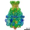


 Sample components
 Processing
 Electron microscopy