 Movie
Movie Controller

[English] 日本語
 Yorodumi
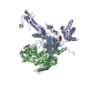
- EMDB-2222: Structure of the PilF DNA transformation ATPase (AMPPNP bound form) -
+ Open data

- Basic information
| Entry | Database: EMDB / ID: EMD-2222 | |||||||||
|---|---|---|---|---|---|---|---|---|---|---|
| Title | Structure of the PilF DNA transformation ATPase (AMPPNP bound form) | |||||||||
 Map data Map data | PilF DNA Transformation ATPase (apoprotein form) | |||||||||
 Sample Sample |
| |||||||||
 Keywords Keywords | ATPase / DNA transformation | |||||||||
| Biological species |   Thermus thermophilus (bacteria) Thermus thermophilus (bacteria) | |||||||||
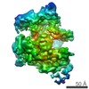
| Method | single particle reconstruction / cryo EM / Resolution: 12.0 Å | |||||||||
 Authors Authors | Collins RF / Hassan D / Karuppiah V / Thistlethwaite A / Derrick JP | |||||||||
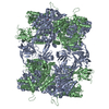
 Citation Citation | Journal: Biochem J / Year: 2013 Title: Structure and mechanism of the PilF DNA transformation ATPase from Thermus thermophilus. Authors: Richard F Collins / Darin Hassan / Vijaykumar Karuppiah / Angela Thistlethwaite / Jeremy P Derrick /  Abstract: Many Gram-negative bacteria contain specific systems for uptake of foreign DNA, which play a critical role in the acquisition of antibiotic resistance. The TtPilF (PilF ATPase from Thermus ...Many Gram-negative bacteria contain specific systems for uptake of foreign DNA, which play a critical role in the acquisition of antibiotic resistance. The TtPilF (PilF ATPase from Thermus thermophilus) is required for high transformation efficiency, but its mechanism of action is unknown. In the present study, we show that TtPilF is able to bind to both DNA and RNA. The structure of TtPilF was determined by cryoelectron microscopy in the presence and absence of the ATP analogue p[NH]ppA (adenosine 5'-[β,γ-imido]triphosphate), at 10 and 12 Å (1 Å=0.1 nm) resolutions respectively. It consists of two distinct N- and C-terminal regions, separated by a short stem-like structure. Binding of p[NH]ppA induces structural changes in the C-terminal domains, which are transmitted via the stem to the N-terminal domains. Molecular models were generated for the apoenzyme and p[NH]ppA-bound states in the C-terminal regions by docking of a model based on a crystal structure from a closely related enzyme. Analysis of DNA binding by electron microscopy, using gold labelling, localized the binding site to the N-terminal domains. The results suggest a model in which DNA uptake by TtPilF is powered by ATP hydrolysis, causing conformational changes in the C-terminal domains, which are transmitted via the stem to take up DNA into the cell. | |||||||||
| History |
|
- Structure visualization
| Movie |
 Movie viewer Movie viewer |
|---|---|
| Structure viewer | EM map: SurfViewMolmilJmol/JSmol |
| Supplemental images |

- Downloads & links
-EMDB archive
| Map data | emd_2222.map.gz | 1.1 MB | EMDB map data format | |
|---|---|---|---|---|
| Header (meta data) | emd-2222-v30.xmlemd-2222.xml | 9 KB 9 KB | Display Display | EMDB header |
| Images |  emd_2222.png emd_2222.png | 106.9 KB | ||
| Archive directory |  http://ftp.pdbj.org/pub/emdb/structures/EMD-2222ftp://ftp.pdbj.org/pub/emdb/structures/EMD-2222 http://ftp.pdbj.org/pub/emdb/structures/EMD-2222ftp://ftp.pdbj.org/pub/emdb/structures/EMD-2222 | HTTPS FTP |
-Validation report
| Summary document | emd_2222_validation.pdf.gz | 200.4 KB | Display | EMDB validaton report |
|---|---|---|---|---|
| Full document | emd_2222_full_validation.pdf.gz | 199.5 KB | Display | |
| Data in XML | emd_2222_validation.xml.gz | 5.1 KB | Display | |
| Arichive directory | https://ftp.pdbj.org/pub/emdb/validation_reports/EMD-2222ftp://ftp.pdbj.org/pub/emdb/validation_reports/EMD-2222 | HTTPS FTP |
-Related structure data


-Links
| EMDB pages | EMDB (EBI/PDBe) / EMDataResource |
|---|
-Map
| File | Download / File: emd_2222.map.gz / Format: CCP4 / Size: 7.8 MB / Type: IMAGE STORED AS FLOATING POINT NUMBER (4 BYTES) | ||||||||||||||||||||||||||||||||||||||||||||||||||||||||||||||||||||
|---|---|---|---|---|---|---|---|---|---|---|---|---|---|---|---|---|---|---|---|---|---|---|---|---|---|---|---|---|---|---|---|---|---|---|---|---|---|---|---|---|---|---|---|---|---|---|---|---|---|---|---|---|---|---|---|---|---|---|---|---|---|---|---|---|---|---|---|---|---|
| Annotation | PilF DNA Transformation ATPase (apoprotein form) | ||||||||||||||||||||||||||||||||||||||||||||||||||||||||||||||||||||
| Voxel size | X=Y=Z: 3 Å | ||||||||||||||||||||||||||||||||||||||||||||||||||||||||||||||||||||
| Density |
| ||||||||||||||||||||||||||||||||||||||||||||||||||||||||||||||||||||
| Symmetry | Space group: 1 | ||||||||||||||||||||||||||||||||||||||||||||||||||||||||||||||||||||
| Details | EMDB XML:
CCP4 map header:
| ||||||||||||||||||||||||||||||||||||||||||||||||||||||||||||||||||||
-Supplemental data
- Sample components
-Entire : PilF ATPase from Thermus thermophilus
| Entire | Name: PilF ATPase from Thermus thermophilus |
|---|---|
| Components |
|
-Supramolecule #1000: PilF ATPase from Thermus thermophilus
| Supramolecule | Name: PilF ATPase from Thermus thermophilus / type: sample / ID: 1000 / Details: Monodisperse sample / Oligomeric state: homohexamer / Number unique components: 1 |
|---|---|
| Molecular weight | Theoretical: 586 KDa |
-Macromolecule #1: ATPase
| Macromolecule | Name: ATPase / type: protein_or_peptide / ID: 1 / Number of copies: 6 / Oligomeric state: Hexamer / Recombinant expression: No |
|---|---|
| Source (natural) | Organism: Thermus thermophilus (bacteria) |
| Molecular weight | Theoretical: 586 KDa |
-Experimental details
-Structure determination
| Method | cryo EM |
|---|---|
 Processing Processing | single particle reconstruction |
| Aggregation state | particle |
-Sample preparation
| Concentration | 0.2 mg/mL |
|---|---|
| Buffer | pH: 6.5 / Details: 50mM Mes pH 6.5, 200mM NaCl and 10mM MgCl2 |
| Grid | Details: Freshly glow discharged 2-2 Quantifoil grids |
| Vitrification | Cryogen name: ETHANE / Chamber humidity: 90 % / Instrument: FEI VITROBOT MARK IV Method: continuously blotted for 4-5 seconds in a 90% humidity chamber before plunge freezing into liquid ethane at 22 degrees. |
- Electron microscopy
| Microscope | FEI POLARA 300 |
|---|---|
| Temperature | Average: 85 K |
| Specialist optics | Energy filter - Name: FEI |
| Date | Nov 1, 2011 |
| Image recording | Digitization - Sampling interval: 3.5 µm / Number real images: 97 / Average electron dose: 20 e/Å2 / Details: Data were recorded on a Gatan 4Kx4K CCD / Bits/pixel: 8 |
| Tilt angle min | 0 |
| Tilt angle max | 0 |
| Electron beam | Acceleration voltage: 200 kV / Electron source:  FIELD EMISSION GUN FIELD EMISSION GUN |
| Electron optics | Calibrated magnification: 39000 / Illumination mode: SPOT SCAN / Imaging mode: BRIGHT FIELD / Cs: 2 mm / Nominal defocus max: 5.0 µm / Nominal defocus min: 0.5 µm |
| Sample stage | Specimen holder: Gatan MSC / Specimen holder model: GATAN LIQUID NITROGEN |
| Experimental equipment |  Model: Tecnai Polara / Image courtesy: FEI Company |
-Image processing
| Details | Semi-automated picking as implemented within EMAN2 |
|---|---|
| CTF correction | Details: EMAN2 - each particle |
| Final reconstruction | Applied symmetry - Point group: C6 (6 fold cyclic) / Algorithm: OTHER / Resolution.type: BY AUTHOR / Resolution: 12.0 Å / Resolution method: FSC 0.5 CUT-OFF / Software - Name: EMAN2 / Number images used: 37000 |
