 Movie
Movie Controller

[English] 日本語
 Yorodumi
- EMDB-2180: 3D reconstruction of LDL at 37C (human body temperature) using cr... -
+ Open data

- Basic information
| Entry | Database: EMDB / ID: EMD-2180 | |||||||||
|---|---|---|---|---|---|---|---|---|---|---|
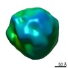
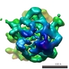
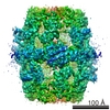
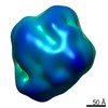
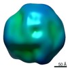
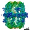
| Title | 3D reconstruction of LDL at 37C (human body temperature) using cryo-EM techniques | |||||||||
 Map data Map data | Reconstruction of native LDL at 37C using cryo-EM based single particle reconstruction | |||||||||
 Sample Sample |
| |||||||||
 Keywords Keywords | LDL / native LDL / human body temperature | |||||||||
| Biological species |   Homo sapiens (human) Homo sapiens (human) | |||||||||
| Method | single particle reconstruction / cryo EM / Resolution: 16.0 Å | |||||||||
 Authors Authors | Kumar V / Butcher SJ / Oorni K / Engelhardt P / Heikkonen J / Kaski K / Ala-Korpela M / Kovanen PT | |||||||||
 Citation Citation | Journal: PLoS One / Year: 2011 Title: Three-dimensional cryoEM reconstruction of native LDL particles to 16Å resolution at physiological body temperature. Authors: Vibhor Kumar / Sarah J Butcher / Katariina Öörni / Peter Engelhardt / Jukka Heikkonen / Kimmo Kaski / Mika Ala-Korpela / Petri T Kovanen /  Abstract: BACKGROUND: Low-density lipoprotein (LDL) particles, the major carriers of cholesterol in the human circulation, have a key role in cholesterol physiology and in the development of atherosclerosis. ...BACKGROUND: Low-density lipoprotein (LDL) particles, the major carriers of cholesterol in the human circulation, have a key role in cholesterol physiology and in the development of atherosclerosis. The most prominent structural components in LDL are the core-forming cholesteryl esters (CE) and the particle-encircling single copy of a huge, non-exchangeable protein, the apolipoprotein B-100 (apoB-100). The shape of native LDL particles and the conformation of native apoB-100 on the particles remain incompletely characterized at the physiological human body temperature (37 °C). METHODOLOGY/PRINCIPAL FINDINGS: To study native LDL particles, we applied cryo-electron microscopy to calculate 3D reconstructions of LDL particles in their hydrated state. Images of the particles ...METHODOLOGY/PRINCIPAL FINDINGS: To study native LDL particles, we applied cryo-electron microscopy to calculate 3D reconstructions of LDL particles in their hydrated state. Images of the particles vitrified at 6 °C and 37 °C resulted in reconstructions at ~16 Å resolution at both temperatures. 3D variance map analysis revealed rigid and flexible domains of lipids and apoB-100 at both temperatures. The reconstructions showed less variability at 6 °C than at 37 °C, which reflected increased order of the core CE molecules, rather than decreased mobility of the apoB-100. Compact molecular packing of the core and order in a lipid-binding domain of apoB-100 were observed at 6 °C, but not at 37 °C. At 37 °C we were able to highlight features in the LDL particles that are not clearly separable in 3D maps at 6 °C. Segmentation of apoB-100 density, fitting of lipovitellin X-ray structure, and antibody mapping, jointly revealed the approximate locations of the individual domains of apoB-100 on the surface of native LDL particles. CONCLUSIONS/SIGNIFICANCE: Our study provides molecular background for further understanding of the link between structure and function of native LDL particles at physiological body temperature. | |||||||||
| History |
|
- Structure visualization
| Movie |
 Movie viewer Movie viewer |
|---|---|
| Structure viewer | EM map: SurfViewMolmilJmol/JSmol |
| Supplemental images |

- Downloads & links
-EMDB archive
| Map data | emd_2180.map.gz | 30.7 MB | EMDB map data format | |
|---|---|---|---|---|
| Header (meta data) | emd-2180-v30.xmlemd-2180.xml | 8.8 KB 8.8 KB | Display Display | EMDB header |
| Images | emd_2180.tif | 914.1 KB | ||
| Archive directory |  http://ftp.pdbj.org/pub/emdb/structures/EMD-2180ftp://ftp.pdbj.org/pub/emdb/structures/EMD-2180 http://ftp.pdbj.org/pub/emdb/structures/EMD-2180ftp://ftp.pdbj.org/pub/emdb/structures/EMD-2180 | HTTPS FTP |
-Related structure data
| Similar structure data |
|---|
-Links
| EMDB pages | EMDB (EBI/PDBe) / EMDataResource |
|---|
-Map
| File | Download / File: emd_2180.map.gz / Format: CCP4 / Size: 33.5 MB / Type: IMAGE STORED AS FLOATING POINT NUMBER (4 BYTES) | ||||||||||||||||||||||||||||||||||||||||||||||||||||||||||||||||||||
|---|---|---|---|---|---|---|---|---|---|---|---|---|---|---|---|---|---|---|---|---|---|---|---|---|---|---|---|---|---|---|---|---|---|---|---|---|---|---|---|---|---|---|---|---|---|---|---|---|---|---|---|---|---|---|---|---|---|---|---|---|---|---|---|---|---|---|---|---|---|
| Annotation | Reconstruction of native LDL at 37C using cryo-EM based single particle reconstruction | ||||||||||||||||||||||||||||||||||||||||||||||||||||||||||||||||||||
| Voxel size | X=Y=Z: 1.4 Å | ||||||||||||||||||||||||||||||||||||||||||||||||||||||||||||||||||||
| Density |
| ||||||||||||||||||||||||||||||||||||||||||||||||||||||||||||||||||||
| Symmetry | Space group: 1 | ||||||||||||||||||||||||||||||||||||||||||||||||||||||||||||||||||||
| Details | EMDB XML:
CCP4 map header:
| ||||||||||||||||||||||||||||||||||||||||||||||||||||||||||||||||||||
-Supplemental data
- Sample components
-Entire : LDL at 37C
| Entire | Name: LDL at 37C |
|---|---|
| Components |
|
-Supramolecule #1000: LDL at 37C
| Supramolecule | Name: LDL at 37C / type: sample / ID: 1000 Details: Samples were vitrified on Quantifoil holey carbon grids (Quantifoil Micro Tools, GmbH) either at 6C or 37C, allowing pre-equilibration of the sample at the desired temperature for at least ...Details: Samples were vitrified on Quantifoil holey carbon grids (Quantifoil Micro Tools, GmbH) either at 6C or 37C, allowing pre-equilibration of the sample at the desired temperature for at least 45 minutes prior to plunging. Number unique components: 1 |
|---|
-Macromolecule #1: apoB-100
| Macromolecule | Name: apoB-100 / type: protein_or_peptide / ID: 1 / Name.synonym: apoB / Recombinant expression: No |
|---|---|
| Source (natural) | Organism: Homo sapiens (human) / synonym: Human / Tissue: Blood |
-Experimental details
-Structure determination
| Method | cryo EM |
|---|---|
 Processing Processing | single particle reconstruction |
| Aggregation state | particle |
-Sample preparation
| Concentration | 0.3 mg/mL |
|---|---|
| Buffer | pH: 7.4 |
| Vitrification | Cryogen name: ETHANE / Instrument: OTHER |
- Electron microscopy
| Microscope | FEI TECNAI F20 |
|---|---|
| Electron beam | Acceleration voltage: 200 kV / Electron source: FIELD EMISSION GUN |
| Electron optics | Illumination mode: FLOOD BEAM / Imaging mode: BRIGHT FIELDBright-field microscopy |
| Sample stage | Specimen holder model: OTHER |
| Date | Dec 30, 2005 |
| Image recording | Digitization - Scanner: ZEISS SCAI / Number real images: 26083 |
| Experimental equipment |  Model: Tecnai F20 / Image courtesy: FEI Company |
-Image processing
| CTF correction | Details: Each particle |
|---|---|
| Final reconstruction | Applied symmetry - Point group: C1 (asymmetric) / Algorithm: OTHER / Resolution.type: BY AUTHOR / Resolution: 16.0 Å / Resolution method: FSC 0.5 CUT-OFF / Software - Name: EMAN / Number images used: 29844 |
| Details | Initially only phase-flipping was carried out to correct for the contrast transfer function. To reduce the effect of image noise and artefacts, an initial single particle reconstruction was made on images de-noised with an information theory [Minimum Description Length (MDL)] based method which we have introduced earlier. Fifty initial class averages were obtained using a reference-free classification of 3000 images by multivariate statistical analysis using parameters, as suggested in EMAN . Angles were assigned using the cross common lines method prior to reconstruction using weighted-back projection. The initial 3D models were low-pass filtered. The 2D image dataset was increased to include all of the particles and iterative refinement continued. After each iteration of the single-particle-reconstruction process only 75% of the images with the highest correlation values were chosen. In order to avoid any bias in 3D reconstruction due to the de-noising, the final reconstructions were made using raw images after being assigned to classes using their de-noised equivalents |
