 Movie
Movie Controller


+ Open data

- Basic information
| Entry | Database: EMDB / ID: EMD-2099 | |||||||||
|---|---|---|---|---|---|---|---|---|---|---|
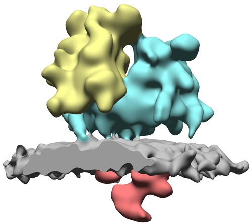
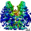
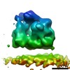
| Title | Structure of ER membrane associated ribosomes in situ | |||||||||
 Map data Map data | Reconstruction of ER membrane associated ribosome from canine pancreatic microsomes | |||||||||
 Sample Sample |
| |||||||||
 Keywords Keywords |  80S ribosome / translocon / mammalian / ER membrane 80S ribosome / translocon / mammalian / ER membrane | |||||||||
| Biological species |  Canis lupus familiaris (dog) Canis lupus familiaris (dog) | |||||||||
| Method | subtomogram averaging / cryo EM / Resolution: 31.0 Å | |||||||||
 Authors Authors | Pfeffer S / Brandt F / Hrabe T / Lang S / Eibauer M / Zimmermann R / Foerster F | |||||||||
 Citation Citation | Journal: Structure / Year: 2012 Title: Structure and 3D arrangement of endoplasmic reticulum membrane-associated ribosomes. Authors: Stefan Pfeffer / Florian Brandt / Thomas Hrabe / Sven Lang / Matthias Eibauer / Richard Zimmermann / Friedrich Förster /  Abstract: In eukaryotic cells, cotranslational protein translocation across the endoplasmic reticulum (ER) membrane requires an elaborate macromolecular machinery. While structural details of ribosomes bound ...In eukaryotic cells, cotranslational protein translocation across the endoplasmic reticulum (ER) membrane requires an elaborate macromolecular machinery. While structural details of ribosomes bound to purified and solubilized constituents of the translocon have been elucidated in recent years, little structural knowledge of ribosomes bound to the complete ER protein translocation machinery in a native membrane environment exists. Here, we used cryoelectron tomography to provide a three-dimensional reconstruction of 80S ribosomes attached to functional canine pancreatic ER microsomes in situ. In the resulting subtomogram average at 31 Å resolution, we observe direct contact of ribosomal expansion segment ES27L and the membrane and distinguish several membrane-embedded and lumenal complexes, including Sec61, the TRAP complex and another large complex protruding 90 Å into the lumen. Membrane-associated ribosomes adopt a preferred three-dimensional arrangement that is likely specific for ER-associated polyribosomes and may explain the high translation efficiency of ER-associated ribosomes compared to their cytosolic counterparts. | |||||||||
| History |
|
- Structure visualization
| Movie |
 Movie viewer Movie viewer |
|---|---|
| Structure viewer | EM map: SurfViewMolmilJmol/JSmol |
| Supplemental images |
- Downloads & links
-EMDB archive
| Map data | emd_2099.map.gz | 7.4 MB | EMDB map data format | |
|---|---|---|---|---|
| Header (meta data) | emd-2099-v30.xmlemd-2099.xml | 9.8 KB 9.8 KB | Display Display | EMDB header |
| Images | EMD-2099-pic.tif | 145.7 KB | ||
| Archive directory |  http://ftp.pdbj.org/pub/emdb/structures/EMD-2099ftp://ftp.pdbj.org/pub/emdb/structures/EMD-2099 http://ftp.pdbj.org/pub/emdb/structures/EMD-2099ftp://ftp.pdbj.org/pub/emdb/structures/EMD-2099 | HTTPS FTP |
-Related structure data
| Similar structure data |
|---|
-Links
| EMDB pages | EMDB (EBI/PDBe) / EMDataResource |
|---|---|
| Related items in Molecule of the Month |

-Map
| File | Download / File: emd_2099.map.gz / Format: CCP4 / Size: 7.8 MB / Type: IMAGE STORED AS FLOATING POINT NUMBER (4 BYTES) | ||||||||||||||||||||||||||||||||||||||||||||||||||||||||||||||||||||
|---|---|---|---|---|---|---|---|---|---|---|---|---|---|---|---|---|---|---|---|---|---|---|---|---|---|---|---|---|---|---|---|---|---|---|---|---|---|---|---|---|---|---|---|---|---|---|---|---|---|---|---|---|---|---|---|---|---|---|---|---|---|---|---|---|---|---|---|---|---|
| Annotation | Reconstruction of ER membrane associated ribosome from canine pancreatic microsomes | ||||||||||||||||||||||||||||||||||||||||||||||||||||||||||||||||||||
| Voxel size | X=Y=Z: 4.68 Å | ||||||||||||||||||||||||||||||||||||||||||||||||||||||||||||||||||||
| Density |
| ||||||||||||||||||||||||||||||||||||||||||||||||||||||||||||||||||||
| Symmetry | Space group: 1 | ||||||||||||||||||||||||||||||||||||||||||||||||||||||||||||||||||||
| Details | EMDB XML:
CCP4 map header:
| ||||||||||||||||||||||||||||||||||||||||||||||||||||||||||||||||||||
-Supplemental data
- Sample components
-Entire : ER membrane associated ribosome
| Entire | Name: ER membrane associated ribosome |
|---|---|
| Components |
|
-Supramolecule #1000: ER membrane associated ribosome
| Supramolecule | Name: ER membrane associated ribosome / type: sample / ID: 1000 / Details: The sample is embedded into its native membrane / Oligomeric state: ER membrane associated ribosome / Number unique components: 1 |
|---|---|
| Molecular weight | Theoretical: 4.5 MDa |
-Supramolecule #1: Membrane-bound 80S ribosome
| Supramolecule | Name: Membrane-bound 80S ribosome / type: complex / ID: 1 / Recombinant expression: No / Database: NCBI / Ribosome-details: ribosome-eukaryote: ALL |
|---|---|
| Source (natural) | Organism: Canis lupus familiaris (dog) / synonym: Dog / Tissue: Pancreas / Organelle: Endoplasmic reticulum / Location in cell: Endoplasmic reticulum |
| Molecular weight | Theoretical: 4.5 MDa |
-Experimental details
-Structure determination
| Method | cryo EM |
|---|---|
 Processing Processing | subtomogram averaging |
| Aggregation state | particle |
-Sample preparation
| Concentration | 1 mg/mL |
|---|---|
| Buffer | pH: 7.4 Details: 5 mM MgCl2, 140 mM KCl, 10 mM Hepes pH 7.4, 1 mM DTT, protease inhibitor |
| Grid | Details: lacy carbon |
| Vitrification | Cryogen name: ETHANE / Instrument: FEI VITROBOT MARK IV / Method: Blot for 3 seconds before plunging |
- Electron microscopy
| Microscope | FEI POLARA 300 |
|---|---|
| Electron beam | Acceleration voltage: 300 kV / Electron source: FIELD EMISSION GUN |
| Electron optics | Illumination mode: FLOOD BEAM / Imaging mode: BRIGHT FIELDBright-field microscopy / Cs: 2 mm / Nominal defocus max: 4.0 µm / Nominal defocus min: 4.0 µm |
| Specialist optics | Energy filter - Name: Gatan GIF 2002 / Energy filter - Lower energy threshold: 0.0 eV / Energy filter - Upper energy threshold: 10.0 eV |
| Sample stage | Specimen holder model: OTHER / Tilt series - Axis1 - Min angle: -60 ° / Tilt series - Axis1 - Max angle: 60 ° |
| Temperature | Min: 80 K / Max: 90 K / Average: 85 K |
| Date | Jan 1, 2011 |
| Image recording | Number real images: 328 / Average electron dose: 50 e/Å2 / Bits/pixel: 12 |
| Experimental equipment |  Model: Tecnai Polara / Image courtesy: FEI Company |
-Image processing
| CTF correction | Details: each particle |
|---|---|
| Final reconstruction | Algorithm: OTHER / Resolution.type: BY AUTHOR / Resolution: 31.0 Å / Resolution method: FSC 0.5 CUT-OFF / Software - Name: PyTom |
| Details | The particles were selected using PyTom and classified by constrained principal component analysis. Average number of projections used in the 3D reconstructions: 1000. Average number of class averages: 1. |
