 Movie
Movie Controller


+ Open data

- Basic information
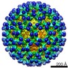
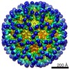
| Entry | Database: EMDB / ID: EMD-2076 | |||||||||
|---|---|---|---|---|---|---|---|---|---|---|
| Title | CryoEM icosahedral reconstruction of AHSV4 | |||||||||
 Map data Map data | AHSV4 filled | |||||||||
 Sample Sample |
| |||||||||
 Keywords Keywords | AHSV4 /  cryoEM / structure cryoEM / structure | |||||||||
| Biological species |  African horse sickness virus 4 African horse sickness virus 4 | |||||||||
| Method | single particle reconstruction / cryo EM / Resolution: 14.4 Å | |||||||||
 Authors Authors | Manole V / Laurinmaki P / Wyngaardt WV / Potgieter CA / Wright IM / Venter GJ / Dijk AAV / Sewell BT / Butcher SJ | |||||||||
 Citation Citation | Journal: J Virol / Year: 2012 Title: Structural insight into African horsesickness virus infection. Authors: Violeta Manole / Pasi Laurinmäki / Wouter Van Wyngaardt / Christiaan A Potgieter / Isabella M Wright / Gert J Venter / Alberdina A van Dijk / B Trevor Sewell / Sarah J Butcher /  Abstract: African horsesickness (AHS) is a devastating disease of horses. The disease is caused by the double-stranded RNA-containing African horsesickness virus (AHSV). Using electron cryomicroscopy and three- ...African horsesickness (AHS) is a devastating disease of horses. The disease is caused by the double-stranded RNA-containing African horsesickness virus (AHSV). Using electron cryomicroscopy and three-dimensional image reconstruction, we determined the architecture of an AHSV serotype 4 (AHSV-4) reference strain. The structure revealed triple-layered AHS virions enclosing the segmented genome and transcriptase complex. The innermost protein layer contains 120 copies of VP3, with the viral polymerase, capping enzyme, and helicase attached to the inner surface of the VP3 layer on the 5-fold axis, surrounded by double-stranded RNA. VP7 trimers form a second, T=13 layer on top of VP3. Comparative analyses of the structures of bluetongue virus and AHSV-4 confirmed that VP5 trimers form globular domains and VP2 trimers form triskelions, on the virion surface. We also identified an AHSV-7 strain with a truncated VP2 protein (AHSV-7 tVP2) which outgrows AHSV-4 in culture. Comparison of AHSV-7 tVP2 to bluetongue virus and AHSV-4 allowed mapping of two domains in AHSV-4 VP2, and one in bluetongue virus VP2, that are important in infection. We also revealed a protein plugging the 5-fold vertices in AHSV-4. These results shed light on virus-host interactions in an economically important orbivirus to help the informed design of new vaccines. | |||||||||
| History |
|
- Structure visualization
| Movie |
 Movie viewer Movie viewer |
|---|---|
| Structure viewer | EM map: SurfViewMolmilJmol/JSmol |
| Supplemental images |

- Downloads & links
-EMDB archive
| Map data | emd_2076.map.gz | 59.9 MB | EMDB map data format | |
|---|---|---|---|---|
| Header (meta data) | emd-2076-v30.xmlemd-2076.xml | 8.2 KB 8.2 KB | Display Display | EMDB header |
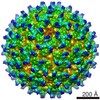
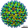
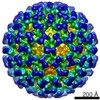
| Images | 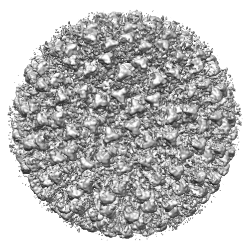 EMD-2076.png EMD-2076.png | 262.8 KB | ||
| Archive directory |  http://ftp.pdbj.org/pub/emdb/structures/EMD-2076ftp://ftp.pdbj.org/pub/emdb/structures/EMD-2076 http://ftp.pdbj.org/pub/emdb/structures/EMD-2076ftp://ftp.pdbj.org/pub/emdb/structures/EMD-2076 | HTTPS FTP |
-Related structure data


-Links
| EMDB pages | EMDB (EBI/PDBe) / EMDataResource |
|---|
-Map
| File | Download / File: emd_2076.map.gz / Format: CCP4 / Size: 120.1 MB / Type: IMAGE STORED AS SIGNED INTEGER (2 BYTES) | ||||||||||||||||||||||||||||||||||||||||||||||||||||||||||||||||||||
|---|---|---|---|---|---|---|---|---|---|---|---|---|---|---|---|---|---|---|---|---|---|---|---|---|---|---|---|---|---|---|---|---|---|---|---|---|---|---|---|---|---|---|---|---|---|---|---|---|---|---|---|---|---|---|---|---|---|---|---|---|---|---|---|---|---|---|---|---|---|
| Annotation | AHSV4 filled | ||||||||||||||||||||||||||||||||||||||||||||||||||||||||||||||||||||
| Voxel size | X=Y=Z: 2.8 Å | ||||||||||||||||||||||||||||||||||||||||||||||||||||||||||||||||||||
| Density |
| ||||||||||||||||||||||||||||||||||||||||||||||||||||||||||||||||||||
| Symmetry | Space group: 1 | ||||||||||||||||||||||||||||||||||||||||||||||||||||||||||||||||||||
| Details | EMDB XML:
CCP4 map header:
| ||||||||||||||||||||||||||||||||||||||||||||||||||||||||||||||||||||
-Supplemental data
- Sample components
-Entire : AHSV4 filled
| Entire | Name: AHSV4 filled |
|---|---|
| Components |
|
-Supramolecule #1000: AHSV4 filled
| Supramolecule | Name: AHSV4 filled / type: sample / ID: 1000 / Number unique components: 1 |
|---|
-Supramolecule #1: African horse sickness virus 4
| Supramolecule | Name: African horse sickness virus 4 / type: virus / ID: 1 / Name.synonym: AHSV4 / NCBI-ID: 36421 / Sci species name: African horse sickness virus 4 / Virus type: VIRION / Virus isolate: SEROTYPE / Virus enveloped: No / Virus empty: No / Syn species name: AHSV4 |
|---|---|
| Host (natural) | Organism:  Equus caballus (horse) / synonym: VERTEBRATES Equus caballus (horse) / synonym: VERTEBRATES |
-Experimental details
-Structure determination
| Method | cryo EM |
|---|---|
 Processing Processing | single particle reconstruction |
| Aggregation state | particle |
-Sample preparation
| Concentration | 0.2 mg/mL |
|---|---|
| Buffer | pH: 8.5 / Details: 2mM Tris-HCl, pH 8.5 |
| Grid | Details: Quantifoil 200 mesh holey carbon |
| Vitrification | Cryogen name: ETHANE / Chamber humidity: 100 % / Instrument: FEI VITROBOT MARK III |
- Electron microscopy
| Microscope | FEI TECNAI F20 |
|---|---|
| Electron beam | Acceleration voltage: 200 kV / Electron source: FIELD EMISSION GUN |
| Electron optics | Calibrated magnification: 50000 / Illumination mode: FLOOD BEAM / Imaging mode: BRIGHT FIELDBright-field microscopy / Cs: 2 mm / Nominal defocus max: 5.0 µm / Nominal defocus min: 0.8 µm / Nominal magnification: 50000 |
| Sample stage | Specimen holder model: GATAN LIQUID NITROGEN |
| Date | Feb 1, 2009 |
| Image recording | Category: FILM / Film or detector model: KODAK SO-163 FILM / Digitization - Scanner: OTHER / Digitization - Sampling interval: 7 µm / Number real images: 307 / Average electron dose: 18 e/Å2 |
| Experimental equipment |  Model: Tecnai F20 / Image courtesy: FEI Company |
-Image processing
| CTF correction | Details: each micrograph |
|---|---|
| Final reconstruction | Applied symmetry - Point group: I (icosahedral) / Algorithm: OTHER / Resolution.type: BY AUTHOR / Resolution: 14.4 Å / Resolution method: FSC 0.5 CUT-OFF / Software - Name: AUTO3DEM / Number images used: 1633 |
