 Movie
Movie Controller


+ Open data

- Basic information
| Entry | Database: EMDB / ID: EMD-2054 | |||||||||
|---|---|---|---|---|---|---|---|---|---|---|
| Title | Electron cryo-microscopy of Mud Crab Reovirus | |||||||||
 Map data Map data | Reconstruction of native Mud Crab Reovirus (MCRV) | |||||||||
 Sample Sample |
| |||||||||
 Keywords Keywords | Mud Crab Reovirus | |||||||||
| Biological species | Mud Crab Reovirus | |||||||||
| Method |  single particle reconstruction / cryo EM / Resolution: 13.8 Å single particle reconstruction / cryo EM / Resolution: 13.8 Å | |||||||||
 Authors Authors | Zengwei H / Xiexiong D / Yinyin L / Hongjun S / Kunpeng L / Zhixun G / Peirui Z / Haidong X / Jianguo H / Qinfen Z / Shaoping W | |||||||||
 Citation Citation | Journal: Virus Res / Year: 2012 Title: Structural insights into the classification of Mud Crab Reovirus. Authors: Zengwei Huang / Xiexiong Deng / Yinyin Li / Hongjun Su / Kunpeng Li / Zhixun Guo / Peirui Zheng / Haidong Xu / Jianguo He / Qinfen Zhang / Shaoping Weng /  Abstract: Cryo-electron microscopy was applied to analyze mud crab reovirus (MCRV), which causes 'sleeping disease' in mud crab, Scylla serrata, a marine species cultured in China. We present here the three ...Cryo-electron microscopy was applied to analyze mud crab reovirus (MCRV), which causes 'sleeping disease' in mud crab, Scylla serrata, a marine species cultured in China. We present here the three dimensional structure of MCRV at 13.8Å resolution. The outer capsid shell is composed of 260 trimers with complete T=13 icosahedral symmetry. A major difference between MCRV and previously reported aquareoviruses is that it lacks a pentameric turret structure. These results together with recently published molecular biological evidence (Deng et al., 2012) indicate that, from a structural perspective, MCRV should be classified as a new member of the family Reoviridae. | |||||||||
| History |
|
- Structure visualization
| Movie |
 Movie viewer Movie viewer |
|---|---|
| Structure viewer | EM map: SurfViewMolmilJmol/JSmol |
| Supplemental images |

- Downloads & links
-EMDB archive
| Map data | emd_2054.map.gz | 31.2 MB | EMDB map data format | |
|---|---|---|---|---|
| Header (meta data) | emd-2054-v30.xmlemd-2054.xml | 9.3 KB 9.3 KB | Display Display | EMDB header |
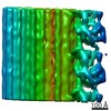
| Images | 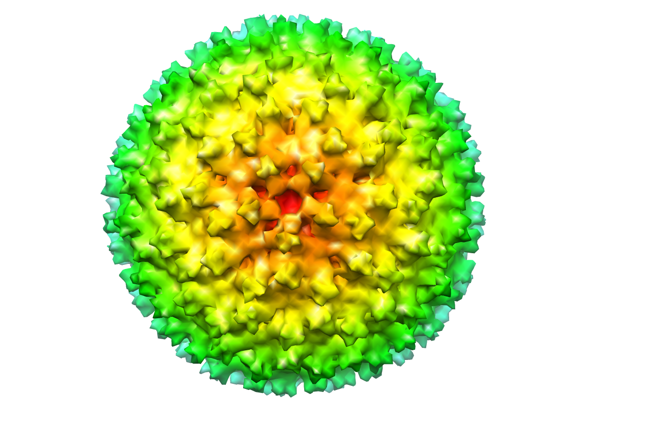 image2054.png image2054.png | 530.2 KB | ||
| Archive directory |  http://ftp.pdbj.org/pub/emdb/structures/EMD-2054ftp://ftp.pdbj.org/pub/emdb/structures/EMD-2054 http://ftp.pdbj.org/pub/emdb/structures/EMD-2054ftp://ftp.pdbj.org/pub/emdb/structures/EMD-2054 | HTTPS FTP |
-Related structure data
| Similar structure data |
|---|


-Links
| EMDB pages | EMDB (EBI/PDBe) / EMDataResource |
|---|
-Map
| File | Download / File: emd_2054.map.gz / Format: CCP4 / Size: 36.7 MB / Type: IMAGE STORED AS FLOATING POINT NUMBER (4 BYTES) | ||||||||||||||||||||||||||||||||||||||||||||||||||||||||||||||||||||
|---|---|---|---|---|---|---|---|---|---|---|---|---|---|---|---|---|---|---|---|---|---|---|---|---|---|---|---|---|---|---|---|---|---|---|---|---|---|---|---|---|---|---|---|---|---|---|---|---|---|---|---|---|---|---|---|---|---|---|---|---|---|---|---|---|---|---|---|---|---|
| Annotation | Reconstruction of native Mud Crab Reovirus (MCRV) | ||||||||||||||||||||||||||||||||||||||||||||||||||||||||||||||||||||
| Voxel size | X=Y=Z: 2.7 Å | ||||||||||||||||||||||||||||||||||||||||||||||||||||||||||||||||||||
| Density |
| ||||||||||||||||||||||||||||||||||||||||||||||||||||||||||||||||||||
| Symmetry | Space group: 1 | ||||||||||||||||||||||||||||||||||||||||||||||||||||||||||||||||||||
| Details | EMDB XML:
CCP4 map header:
| ||||||||||||||||||||||||||||||||||||||||||||||||||||||||||||||||||||
-Supplemental data
- Sample components
-Entire : Mud Crab Reovirus (MCRV)
| Entire | Name: Mud Crab Reovirus (MCRV) |
|---|---|
| Components |
|
-Supramolecule #1000: Mud Crab Reovirus (MCRV)
| Supramolecule | Name: Mud Crab Reovirus (MCRV) / type: sample / ID: 1000 / Oligomeric state: icosahedral / Number unique components: 1 |
|---|
-Supramolecule #1: Mud Crab Reovirus
| Supramolecule | Name: Mud Crab Reovirus / type: virus / ID: 1 / Name.synonym: Mud Crab Reovirus / Sci species name: Mud Crab Reovirus / Virus type: VIRION / Virus isolate: SPECIES / Virus enveloped: No / Virus empty: No / Syn species name: Mud Crab Reovirus |
|---|---|
| Host (natural) | Organism:  Scylla serrata (giant mud crab) / synonym: INVERTEBRATES Scylla serrata (giant mud crab) / synonym: INVERTEBRATES |
| Virus shell | Shell ID: 1 / Diameter: 724 Å / T number (triangulation number): 13 |
-Experimental details
-Structure determination
| Method | cryo EM |
|---|---|
 Processing Processing | single particle reconstruction |
| Aggregation state | particle |
-Sample preparation
| Buffer | pH: 7.2 / Details: 0.1M PBS |
|---|---|
| Grid | Details: R1.2/1.3 Quantifoil grids(200mesh) with thin carbon film |
| Vitrification | Cryogen name: ETHANE / Instrument: HOMEMADE PLUNGER / Details: freezed by manual / Method: blot about 5 seconds by manual before plunging |
- Electron microscopy
| Microscope | JEOL 2010 |
|---|---|
| Electron beam | Acceleration voltage: 200 kV / Electron source: LAB6 |
| Electron optics | Calibrated magnification: 46900 / Illumination mode: FLOOD BEAM / Imaging mode: BRIGHT FIELDBright-field microscopy / Cs: 2.0 mm / Nominal defocus max: 2.0 µm / Nominal defocus min: 1.0 µm / Nominal magnification: 50000 |
| Sample stage | Specimen holder: Liquid nitrogen cooled / Specimen holder model: GATAN LIQUID NITROGEN |
| Temperature | Min: 100 K / Max: 103 K / Average: 102 K |
| Alignment procedure | Legacy - Astigmatism: Objective lens astigmatism was corrected at 400,000 times magnification |
| Details | Miminum dose illumination (MDS model) |
| Date | Jan 12, 2009 |
| Image recording | Category: FILM / Film or detector model: KODAK SO-163 FILM / Digitization - Scanner: NIKON COOLSCAN / Digitization - Sampling interval: 6.5 µm / Number real images: 100 / Average electron dose: 18 e/Å2 / Bits/pixel: 16 |
-Image processing
| CTF correction | Details: Each particle |
|---|---|
| Final reconstruction | Applied symmetry - Point group: I (icosahedral) / Resolution.type: BY AUTHOR / Resolution: 13.8 Å / Resolution method: FSC 0.5 CUT-OFF / Software - Name: EMAN / Number images used: 4020 |
| Details | The particles were selected by EMAN boxer programms. |
