 Movie
Movie Controller


+ Open data

- Basic information
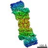
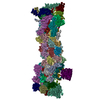
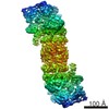
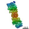
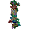
| Entry | Database: EMDB / ID: EMD-2047 | |||||||||
|---|---|---|---|---|---|---|---|---|---|---|
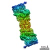
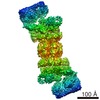
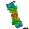
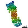
| Title | Structure of the human 26S proteasome | |||||||||
 Map data Map data | The deposited map has been supersampled to a grid of 1.412 angstroms (from the experimental 2.824 angstroms) for improved display | |||||||||
 Sample Sample |
| |||||||||
 Keywords Keywords | 26S proteasome / proteasome / 20S / 19S / human | |||||||||
| Biological species |  Homo sapiens (human) Homo sapiens (human) | |||||||||
| Method | single particle reconstruction / cryo EM / Resolution: 7.0 Å | |||||||||
 Authors Authors | da Fonseca PCA / He J / Morris EP | |||||||||
 Citation Citation | Journal: Mol Cell / Year: 2012 Title: Molecular model of the human 26S proteasome. Authors: Paula C A da Fonseca / Jun He / Edward P Morris /  Abstract: The 26S proteasome plays a fundamental role in eukaryotic homeostasis by undertaking the highly controlled degradation of a wide range of proteins, including key cellular regulators such as those ...The 26S proteasome plays a fundamental role in eukaryotic homeostasis by undertaking the highly controlled degradation of a wide range of proteins, including key cellular regulators such as those controlling cell-cycle progression and apoptosis. Here we report the structure of the human 26S proteasome determined by cryo-electron microscopy and single-particle analysis, with secondary structure elements identified both in the 20S proteolytic core region and in the 19S regulatory particle. We have used this information together with crystal structures, homology models, and other biochemical information to construct a molecular model of the complete 26S proteasome. This model allows for a detailed description of the 20S core within the 26S proteasome and redefines the overall assignment of subunits within the 19S regulatory particle. The information presented here provides a strong basis for a mechanistic understanding of the 26S proteasome. | |||||||||
| History |
|
- Structure visualization
| Movie |
 Movie viewer Movie viewer |
|---|---|
| Structure viewer | EM map: SurfViewMolmilJmol/JSmol |
| Supplemental images |

- Downloads & links
-EMDB archive
| Map data | emd_2047.map.gz | 34.5 MB | EMDB map data format | |
|---|---|---|---|---|
| Header (meta data) | emd-2047-v30.xmlemd-2047.xml | 7.8 KB 7.8 KB | Display Display | EMDB header |
| Images |  EMDEP_2047.jpg EMDEP_2047.jpg | 59.1 KB | ||
| Archive directory |  http://ftp.pdbj.org/pub/emdb/structures/EMD-2047ftp://ftp.pdbj.org/pub/emdb/structures/EMD-2047 http://ftp.pdbj.org/pub/emdb/structures/EMD-2047ftp://ftp.pdbj.org/pub/emdb/structures/EMD-2047 | HTTPS FTP |
-Validation report
| Summary document | emd_2047_validation.pdf.gz | 203.9 KB | Display | EMDB validaton report |
|---|---|---|---|---|
| Full document | emd_2047_full_validation.pdf.gz | 203 KB | Display | |
| Data in XML | emd_2047_validation.xml.gz | 4.6 KB | Display | |
| Arichive directory | https://ftp.pdbj.org/pub/emdb/validation_reports/EMD-2047ftp://ftp.pdbj.org/pub/emdb/validation_reports/EMD-2047 | HTTPS FTP |
-Related structure data
| Similar structure data |
|---|

-Links
| EMDB pages | EMDB (EBI/PDBe) / EMDataResource |
|---|
-Map
| File | Download / File: emd_2047.map.gz / Format: CCP4 / Size: 42.9 MB / Type: IMAGE STORED AS FLOATING POINT NUMBER (4 BYTES) | ||||||||||||||||||||||||||||||||||||||||||||||||||||||||||||||||||||
|---|---|---|---|---|---|---|---|---|---|---|---|---|---|---|---|---|---|---|---|---|---|---|---|---|---|---|---|---|---|---|---|---|---|---|---|---|---|---|---|---|---|---|---|---|---|---|---|---|---|---|---|---|---|---|---|---|---|---|---|---|---|---|---|---|---|---|---|---|---|
| Annotation | The deposited map has been supersampled to a grid of 1.412 angstroms (from the experimental 2.824 angstroms) for improved display | ||||||||||||||||||||||||||||||||||||||||||||||||||||||||||||||||||||
| Voxel size | X=Y=Z: 1.412 Å | ||||||||||||||||||||||||||||||||||||||||||||||||||||||||||||||||||||
| Density |
| ||||||||||||||||||||||||||||||||||||||||||||||||||||||||||||||||||||
| Symmetry | Space group: 1 | ||||||||||||||||||||||||||||||||||||||||||||||||||||||||||||||||||||
| Details | EMDB XML:
CCP4 map header:
| ||||||||||||||||||||||||||||||||||||||||||||||||||||||||||||||||||||
-Supplemental data
- Sample components
-Entire : 26S proteasome
| Entire | Name: 26S proteasome |
|---|---|
| Components |
|
-Supramolecule #1000: 26S proteasome
| Supramolecule | Name: 26S proteasome / type: sample / ID: 1000 / Oligomeric state: dimer / Number unique components: 1 |
|---|---|
| Molecular weight | Theoretical: 2.6 MDa |
-Macromolecule #1: 26S proteasome
| Macromolecule | Name: 26S proteasome / type: protein_or_peptide / ID: 1 / Oligomeric state: dimer / Recombinant expression: No |
|---|---|
| Source (natural) | Organism: Homo sapiens (human) / synonym: Human / Cell: Erythrocyte |
| Molecular weight | Theoretical: 2.6 MDa |
-Experimental details
-Structure determination
| Method | cryo EM |
|---|---|
 Processing Processing | single particle reconstruction |
| Aggregation state | particle |
-Sample preparation
| Concentration | 0.15 mg/mL |
|---|---|
| Buffer | Details: 50 mM Tris-HCl, pH 7.5, mM MgCl2, 2 mM ATP and 1 mM dithiotreitol |
| Grid | Details: Quantifoil coated with thin layer of carbon |
| Vitrification | Cryogen name: ETHANE / Chamber humidity: 90 % / Instrument: FEI VITROBOT MARK III / Method: Blot for 5 seconds |
- Electron microscopy
| Microscope | FEI TECNAI F20 |
|---|---|
| Details | CCD images binned x2 |
| Date | Oct 20, 2010 |
| Image recording | Category: CCD / Film or detector model: TVIPS TEMCAM-F415 (4k x 4k) / Average electron dose: 25 e/Å2 / Bits/pixel: 16 |
| Electron beam | Acceleration voltage: 200 kV / Electron source:  FIELD EMISSION GUN FIELD EMISSION GUN |
| Electron optics | Illumination mode: FLOOD BEAM / Imaging mode: BRIGHT FIELD / Nominal defocus max: 2.475 µm / Nominal defocus min: 1.1 µm / Nominal magnification: 63000 |
| Sample stage | Specimen holder model: GATAN LIQUID NITROGEN |
| Experimental equipment |  Model: Tecnai F20 / Image courtesy: FEI Company |
-Image processing
| CTF correction | Details: CCD frame |
|---|---|
| Final reconstruction | Applied symmetry - Point group: C2 (2 fold cyclic) / Algorithm: OTHER / Resolution.type: BY AUTHOR / Resolution: 7.0 Å / Resolution method: OTHER / Software - Name: IMAGIC,Spider,in-house,software / Number images used: 12718 |
