Movie
Movie Controller

[English] 日本語
 Yorodumi
- EMDB-2046: Cryo-electron microscopy study of hepatitis B virus decorated wit... -
+ Open data

- Basic information
| Entry | Database: EMDB / ID: EMD-2046 | |||||||||
|---|---|---|---|---|---|---|---|---|---|---|
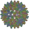
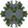
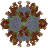
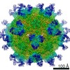
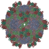
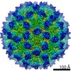
| Title | Cryo-electron microscopy study of hepatitis B virus decorated with the antibody E1 | |||||||||
 Map data Map data | 3D reconstruction of antibody-decorated Hepatitis B T=4 particle | |||||||||
 Sample Sample |
| |||||||||
 Keywords Keywords | Hepatitis B / core antigen / ALF / acute liver failure / antibody E1 / monoclonal / conformational epitope / fulminant hepatitis / cryo-EM | |||||||||
| Biological species |  Homo sapiens (human) / Homo sapiens (human) /   Hepatitis B virus Hepatitis B virus | |||||||||
| Method | single particle reconstruction / cryo EM / Resolution: 10.0 Å | |||||||||
 Authors Authors | Wu W / Chen ZC / Cheng NQ / Watts NR / Stahl SJ / Farci P / Purcell RH / Wingfield PT / Steven AC | |||||||||
 Citation Citation | Journal: J Struct Biol / Year: 2013 Title: Specificity of an anti-capsid antibody associated with Hepatitis B Virus-related acute liver failure. Authors: Weimin Wu / Zhaochun Chen / Naiqian Cheng / Norman R Watts / Stephen J Stahl / Patrizia Farci / Robert H Purcell / Paul T Wingfield / Alasdair C Steven /  Abstract: Previously, the livers of patients suffering from acute liver failure (ALF), a potentially fatal syndrome arising from infection by Hepatitis B Virus (HBV), were found to contain massive amounts of ...Previously, the livers of patients suffering from acute liver failure (ALF), a potentially fatal syndrome arising from infection by Hepatitis B Virus (HBV), were found to contain massive amounts of an antibody specific for the core antigen (HBcAg) capsid. We have used cryo-electron microscopy and molecular modeling to define its epitope. HBV capsids are icosahedral shells with 25Å-long dimeric spikes, each a 4-helix bundle, protruding from the contiguous "floor". Of the anti-HBcAg antibodies previously characterized, most bind around the spike tip while one binds to the floor. The ALF-associated antibody binds tangentially to a novel site on the side of the spike. This epitope is conformational. The Fab binds with high affinity to its principal determinants but has lower affinities for quasi-equivalent variants. The highest occupancy site is on one side of a spike, with no detectable binding to the corresponding site on the other side. Binding of one Fab per dimer was also observed by analytical ultracentrifugation. The Fab did not bind to the e-antigen dimer, a non-assembling variant of capsid protein. These findings support the propositions that antibodies with particular specificities may correlate with different clinical expressions of HBV infection and that antibodies directed to particular HBcAg epitopes may be involved in ALF pathogenesis. | |||||||||
| History |
|
- Structure visualization
| Movie |
 Movie viewer Movie viewer |
|---|---|
| Structure viewer | EM map: SurfViewMolmilJmol/JSmol |
| Supplemental images |
- Downloads & links
-EMDB archive
| Map data | emd_2046.map.gz | 21 MB | EMDB map data format | |
|---|---|---|---|---|
| Header (meta data) | emd-2046-v30.xmlemd-2046.xml | 11.1 KB 11.1 KB | Display Display | EMDB header |
| Images | EMD_2046_icon.tif | 762.9 KB | ||
| Archive directory |  http://ftp.pdbj.org/pub/emdb/structures/EMD-2046ftp://ftp.pdbj.org/pub/emdb/structures/EMD-2046 http://ftp.pdbj.org/pub/emdb/structures/EMD-2046ftp://ftp.pdbj.org/pub/emdb/structures/EMD-2046 | HTTPS FTP |
-Validation report
| Summary document | emd_2046_validation.pdf.gz | 265.1 KB | Display | EMDB validaton report |
|---|---|---|---|---|
| Full document | emd_2046_full_validation.pdf.gz | 264.2 KB | Display | |
| Data in XML | emd_2046_validation.xml.gz | 6.2 KB | Display | |
| Arichive directory | https://ftp.pdbj.org/pub/emdb/validation_reports/EMD-2046ftp://ftp.pdbj.org/pub/emdb/validation_reports/EMD-2046 | HTTPS FTP |
-Related structure data
| Similar structure data |
|---|
-Links
| EMDB pages | EMDB (EBI/PDBe) / EMDataResource |
|---|
-Map
| File | Download / File: emd_2046.map.gz / Format: CCP4 / Size: 29.8 MB / Type: IMAGE STORED AS FLOATING POINT NUMBER (4 BYTES) | ||||||||||||||||||||||||||||||||||||||||||||||||||||||||||||||||||||
|---|---|---|---|---|---|---|---|---|---|---|---|---|---|---|---|---|---|---|---|---|---|---|---|---|---|---|---|---|---|---|---|---|---|---|---|---|---|---|---|---|---|---|---|---|---|---|---|---|---|---|---|---|---|---|---|---|---|---|---|---|---|---|---|---|---|---|---|---|---|
| Annotation | 3D reconstruction of antibody-decorated Hepatitis B T=4 particle | ||||||||||||||||||||||||||||||||||||||||||||||||||||||||||||||||||||
| Voxel size | X=Y=Z: 2.457 Å | ||||||||||||||||||||||||||||||||||||||||||||||||||||||||||||||||||||
| Density |
| ||||||||||||||||||||||||||||||||||||||||||||||||||||||||||||||||||||
| Symmetry | Space group: 1 | ||||||||||||||||||||||||||||||||||||||||||||||||||||||||||||||||||||
| Details | EMDB XML:
CCP4 map header:
| ||||||||||||||||||||||||||||||||||||||||||||||||||||||||||||||||||||
-Supplemental data
- Sample components
-Entire : Antibody E1 to Hepatitis B core antigen
| Entire | Name: Antibody E1 to Hepatitis B core antigen |
|---|---|
| Components |
|
-Supramolecule #1000: Antibody E1 to Hepatitis B core antigen
| Supramolecule | Name: Antibody E1 to Hepatitis B core antigen / type: sample / ID: 1000 Details: The antibody E1 is derived from two ALF patients. T=4 hepatitis B particle is Cp149.3CA Oligomeric state: One antibody to one face of the dimer / Number unique components: 1 |
|---|---|
| Molecular weight | Theoretical: 4.06 MDa |
-Supramolecule #1: Hepatitis B virus
| Supramolecule | Name: Hepatitis B virus / type: virus / ID: 1 / NCBI-ID: 10407 / Sci species name: Hepatitis B virus / Virus type: VIRUS-LIKE PARTICLE / Virus isolate: SPECIES / Virus enveloped: Yes / Virus empty: Yes |
|---|---|
| Host (natural) | Organism: Homo sapiens (human) / synonym: VERTEBRATES |
-Macromolecule #1: Antibody E1
| Macromolecule | Name: Antibody E1 / type: protein_or_peptide / ID: 1 / Details: Molecular weight is for one copy / Recombinant expression: Yes |
|---|---|
| Source (natural) | Organism: Homo sapiens (human) / synonym: Human / Tissue: Plasma / Cell: B-lyphocytes |
| Molecular weight | Theoretical: 50 KDa |
-Experimental details
-Structure determination
| Method | cryo EM |
|---|---|
 Processing Processing | single particle reconstruction |
| Aggregation state | particle |
-Sample preparation
| Vitrification | Cryogen name: ETHANE / Chamber humidity: 38 % / Chamber temperature: 100 K / Instrument: OTHER / Method: Manual plunging |
|---|
- Electron microscopy
| Microscope | FEI/PHILIPS CM200FEG |
|---|---|
| Temperature | Average: 100 K |
| Alignment procedure | Legacy - Astigmatism: Objective lens astigmatism was corrected at 175,000 magnification |
| Date | Jul 1, 2010 |
| Image recording | Category: FILM / Film or detector model: KODAK SO-163 FILM / Digitization - Scanner: NIKON SUPER COOLSCAN 9000 / Digitization - Sampling interval: 6.35 µm / Number real images: 52 / Average electron dose: 20 e/Å2 Details: There are two kinds of particle in each micrograph. T=4 and T=3 Hepatitis B particles. Bits/pixel: 16 |
| Electron beam | Acceleration voltage: 120 kV / Electron source:  FIELD EMISSION GUN FIELD EMISSION GUN |
| Electron optics | Calibrated magnification: 51689 / Illumination mode: FLOOD BEAM / Imaging mode: BRIGHT FIELD / Cs: 2.0 mm / Nominal defocus max: 0.0018 µm / Nominal defocus min: 0.001 µm / Nominal magnification: 5000 |
| Sample stage | Specimen holder model: GATAN LIQUID NITROGEN |
-Image processing
| Details | EMAN and EMAN2 were applied to solve the structure. |
|---|---|
| CTF correction | Details: Micrograph |
| Final reconstruction | Applied symmetry - Point group: I (icosahedral) / Algorithm: OTHER / Resolution.type: BY AUTHOR / Resolution: 10.0 Å / Resolution method: FSC 0.5 CUT-OFF / Software - Name: EMAN,EMAN2 / Number images used: 3787 |
| Final angle assignment | Details: EMAN convention |
-Atomic model buiding 1
| Initial model | PDB ID: Chain - #0 - Chain ID: A / Chain - #1 - Chain ID: B / Chain - #2 - Chain ID: C / Chain - #3 - Chain ID: D |
|---|---|
| Software | Name: Chimera,Situs |
| Details | Protocol: Rigid body. The PDB structure was manually docked into the reconstruction using Chimera and was automatically fitted using Situs. |
| Refinement | Space: REAL / Protocol: RIGID BODY FIT |