 Movie
Movie Controller

[English] 日本語
 Yorodumi
- EMDB-1995: Electron density map of a composite coiled-coil fibril comprising... -
+ Open data

- Basic information
| Entry | Database: EMDB / ID: EMD-1995 | |||||||||
|---|---|---|---|---|---|---|---|---|---|---|
| Title | Electron density map of a composite coiled-coil fibril comprising multiple self-assembling fibre peptides. | |||||||||
 Map data Map data | This is a map of a coiled coil derived from a micrograph of a 3D protein fibre processed using 2dx | |||||||||
 Sample Sample |
| |||||||||
 Keywords Keywords | CryoTEM /  coiled coil / fibrous proteins / protein design / self-assembly coiled coil / fibrous proteins / protein design / self-assembly | |||||||||
| Biological species | synthetic construct (others) | |||||||||
| Method | electron crystallography / cryo EM / Resolution: 8.0 Å | |||||||||
 Authors Authors | Sharp TH / Bruning M / Mantell J / Sessions RB / Thomson AR / Zaccai NR / Brady RL / Verkade P / Woolfson DN | |||||||||
 Citation Citation | Journal: Proc Natl Acad Sci U S A / Year: 2012 Title: Cryo-transmission electron microscopy structure of a gigadalton peptide fiber of de novo design. Authors: Thomas H Sharp / Marc Bruning / Judith Mantell / Richard B Sessions / Andrew R Thomson / Nathan R Zaccai / R Leo Brady / Paul Verkade / Derek N Woolfson /  Abstract: Nature presents various protein fibers that bridge the nanometer to micrometer regimes. These structures provide inspiration for the de novo design of biomimetic assemblies, both to address ...Nature presents various protein fibers that bridge the nanometer to micrometer regimes. These structures provide inspiration for the de novo design of biomimetic assemblies, both to address difficulties in studying and understanding natural systems, and to provide routes to new biomaterials with potential applications in nanotechnology and medicine. We have designed a self-assembling fiber system, the SAFs, in which two small α-helical peptides are programmed to form a dimeric coiled coil and assemble in a controlled manner. The resulting fibers are tens of nm wide and tens of μm long, and, therefore, comprise millions of peptides to give gigadalton supramolecular structures. Here, we describe the structure of the SAFs determined to approximately 8 Å resolution using cryotransmission electron microscopy. Individual micrographs show clear ultrastructure that allowed direct interpretation of the packing of individual α-helices within the fibers, and the construction of a 3D electron density map. Furthermore, a model was derived using the cryotransmission electron microscopy data and side chains taken from a 2.3 Å X-ray crystal structure of a peptide building block incapable of forming fibers. This was validated using single-particle analysis techniques, and was stable in prolonged molecular-dynamics simulation, confirming its structural viability. The level of self-assembly and self-organization in the SAFs is unprecedented for a designed peptide-based material, particularly for a system of considerably reduced complexity compared with natural proteins. This structural insight is a unique high-resolution description of how α-helical fibrils pack into larger protein fibers, and provides a basis for the design and engineering of future biomaterials. | |||||||||
| History |
|
- Structure visualization
| Movie |
 Movie viewer Movie viewer |
|---|---|
| Structure viewer | EM map: SurfViewMolmilJmol/JSmol |
| Supplemental images |

- Downloads & links
-EMDB archive
| Map data | emd_1995.map.gz | 3.6 MB | EMDB map data format | |
|---|---|---|---|---|
| Header (meta data) | emd-1995-v30.xmlemd-1995.xml | 11.5 KB 11.5 KB | Display Display | EMDB header |
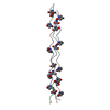
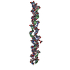
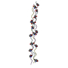
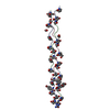
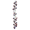
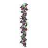
| Images | 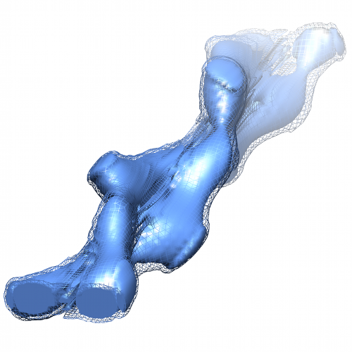 SAF.png SAF.png | 243.3 KB | ||
| Archive directory |  http://ftp.pdbj.org/pub/emdb/structures/EMD-1995ftp://ftp.pdbj.org/pub/emdb/structures/EMD-1995 http://ftp.pdbj.org/pub/emdb/structures/EMD-1995ftp://ftp.pdbj.org/pub/emdb/structures/EMD-1995 | HTTPS FTP |
-Related structure data

-Links
| EMDB pages | EMDB (EBI/PDBe) / EMDataResource |
|---|
-Map
| File | Download / File: emd_1995.map.gz / Format: CCP4 / Size: 4.8 MB / Type: IMAGE STORED AS FLOATING POINT NUMBER (4 BYTES) | ||||||||||||||||||||||||||||||||||||||||||||||||||||||||||||||||||||
|---|---|---|---|---|---|---|---|---|---|---|---|---|---|---|---|---|---|---|---|---|---|---|---|---|---|---|---|---|---|---|---|---|---|---|---|---|---|---|---|---|---|---|---|---|---|---|---|---|---|---|---|---|---|---|---|---|---|---|---|---|---|---|---|---|---|---|---|---|---|
| Annotation | This is a map of a coiled coil derived from a micrograph of a 3D protein fibre processed using 2dx | ||||||||||||||||||||||||||||||||||||||||||||||||||||||||||||||||||||
| Voxel size | X=Y=Z: 0.627 Å | ||||||||||||||||||||||||||||||||||||||||||||||||||||||||||||||||||||
| Density |
| ||||||||||||||||||||||||||||||||||||||||||||||||||||||||||||||||||||
| Symmetry | Space group: 1 | ||||||||||||||||||||||||||||||||||||||||||||||||||||||||||||||||||||
| Details | EMDB XML:
CCP4 map header:
| ||||||||||||||||||||||||||||||||||||||||||||||||||||||||||||||||||||
-Supplemental data
- Sample components
-Entire : Self-assembling peptide fibre
| Entire | Name: Self-assembling peptide fibre |
|---|---|
| Components |
|
-Supramolecule #1000: Self-assembling peptide fibre
| Supramolecule | Name: Self-assembling peptide fibre / type: sample / ID: 1000 / Details: Samples contains approximately 30,000,000 peptides Oligomeric state: Many millions of peptides self-assemble to form a gigadalton peptide fibre Number unique components: 2 |
|---|
-Macromolecule #1: KIAALKQKIASLKQEIDALEYENDALEQ
| Macromolecule | Name: KIAALKQKIASLKQEIDALEYENDALEQ / type: protein_or_peptide / ID: 1 / Name.synonym: Self-assembling fibre Details: Peptides were synthesized by microwave-assisted solid-phase peptide synthesis using standard HBTU activation. They heterodimerize to form offset dimeric coiled coils with complementary ...Details: Peptides were synthesized by microwave-assisted solid-phase peptide synthesis using standard HBTU activation. They heterodimerize to form offset dimeric coiled coils with complementary sticky ends that assemble to form extended coiled coil fibrils. These fibrils pack laterally to generate large proteinaceous fibres on average 82 nm in diameter and 42 micrometers in length. Recombinant expression: No / Database: NCBI |
|---|---|
| Source (natural) | Organism: synthetic construct (others) |
| Molecular weight | Theoretical: 3.17 KDa |
-Macromolecule #2: KIRRLKQKNARLKQEIAALEYEIAALEQ
| Macromolecule | Name: KIRRLKQKNARLKQEIAALEYEIAALEQ / type: protein_or_peptide / ID: 2 / Name.synonym: Self-assembling fibre Details: Peptides were synthesized by microwave-assisted solid-phase peptide synthesis using standard HBTU activation. They heterodimerize to form offset dimeric coiled coils with complementary ...Details: Peptides were synthesized by microwave-assisted solid-phase peptide synthesis using standard HBTU activation. They heterodimerize to form offset dimeric coiled coils with complementary sticky ends that assemble to form extended coiled coil fibrils. These fibrils pack laterally to generate large proteinaceous fibres on average 82 nm in diameter and 42 micrometers in length. Recombinant expression: No / Database: NCBI |
|---|---|
| Source (natural) | Organism: synthetic construct (others) |
| Molecular weight | Theoretical: 3.32 KDa |
-Experimental details
-Structure determination
| Method | cryo EM |
|---|---|
 Processing Processing | electron crystallography |
| Aggregation state | 2D array |
-Sample preparation
| Concentration | 0.325 mg/mL |
|---|---|
| Buffer | pH: 7.4 / Details: 10 mM MOPS |
| Grid | Details: Lacey-carbon grid |
| Vitrification | Cryogen name: ETHANE / Chamber humidity: 100 % / Chamber temperature: 98 K / Instrument: OTHER / Details: Vitrification instrument: Vitrobot / Method: Blot 1 sec. |
- Electron microscopy
| Microscope | FEI TECNAI 20 |
|---|---|
| Electron beam | Acceleration voltage: 200 kV / Electron source: LAB6 |
| Electron optics | Illumination mode: FLOOD BEAM / Imaging mode: BRIGHT FIELDBright-field microscopy / Cs: 2 mm / Nominal defocus max: 1.5 µm / Nominal defocus min: 1.5 µm / Nominal magnification: 50000 |
| Sample stage | Specimen holder: liquid nitrogen-cooled cryo specimen holder Specimen holder model: GATAN LIQUID NITROGEN / Tilt series - Axis1 - Min angle: 0 ° / Tilt series - Axis1 - Max angle: 0 ° |
| Details | Low dose software used |
| Date | Apr 20, 2010 |
| Image recording | Digitization - Sampling interval: 11 µm / Number real images: 1 / Average electron dose: 10 e/Å2 |
| Tilt angle min | 0 |
| Tilt angle max | 0 |
-Image processing
| Crystal parameters | Unit cell - A: 20.8 Å / Unit cell - B: 20.8 Å / Unit cell - C: 125.4 Å / Unit cell - γ: 120 ° / Unit cell - α: 90 ° / Unit cell - β: 90 ° / Plane group: P 1 |
|---|---|
| CTF correction | Details: Each image |
| Final reconstruction | Algorithm: OTHER / Resolution.type: BY AUTHOR / Resolution: 8.0 Å / Resolution method: OTHER / Software - Name: 2dx, BHP |
