Movie
Movie Controller

[English] 日本語
 Yorodumi
- EMDB-1985: Structure of the full human RXR-VDR nuclear receptor heterodimer ... -
+ Open data

- Basic information
| Entry | Database: EMDB / ID: EMD-1985 | |||||||||
|---|---|---|---|---|---|---|---|---|---|---|
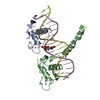
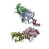
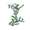
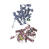
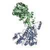
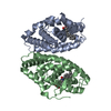
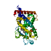
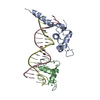
| Title | Structure of the full human RXR-VDR nuclear receptor heterodimer complex with its DR3 target DNA | |||||||||
 Map data Map data | VDR-RXR DR3 DNA complex | |||||||||
 Sample Sample |
| |||||||||
 Keywords Keywords |  Nuclear receptor / retinoic acid / vitamin D / DNA response element Nuclear receptor / retinoic acid / vitamin D / DNA response element | |||||||||
| Biological species |  Homo sapiens (human) Homo sapiens (human) | |||||||||
| Method | single particle reconstruction / cryo EM / negative staining / Resolution: 12.0 Å | |||||||||
 Authors Authors | Orlov I / Rochel N / Moras D / Klaholz BP | |||||||||
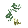
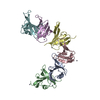
 Citation Citation | Journal: EMBO J / Year: 2012 Title: Structure of the full human RXR/VDR nuclear receptor heterodimer complex with its DR3 target DNA. Authors: Igor Orlov / Natacha Rochel / Dino Moras / Bruno P Klaholz /  Abstract: Transcription regulation by steroid hormones and other metabolites is mediated by nuclear receptors (NRs) such as the vitamin D and retinoid X receptors (VDR and RXR). Here, we present the cryo ...Transcription regulation by steroid hormones and other metabolites is mediated by nuclear receptors (NRs) such as the vitamin D and retinoid X receptors (VDR and RXR). Here, we present the cryo electron microscopy (cryo-EM) structure of the heterodimeric complex of the liganded human RXR and VDR bound to a consensus DNA response element forming a direct repeat (DR3). The cryo-EM map of the 100-kDa complex allows positioning the individual crystal structures of ligand- and DNA-binding domains (LBDs and DBDs). The LBDs are arranged perpendicular to the DNA and are located asymmetrically at the DNA 5'-end of the response element. The structure reveals that the VDR N-terminal A/B domain is located close to the DNA. The hinges of both VDR and RXR are fully visible and hold the complex in an open conformation in which co-regulators can bind. The asymmetric topology of the complex provides the structural basis for RXR being an adaptive partner within NR heterodimers, while the specific helical structure of VDR's hinge connects the 3'-bound DBD with the 5'-bound LBD and thereby serves as a conserved linker of defined length sensitive to mutational deletion. | |||||||||
| History |
|
- Structure visualization
| Movie |
 Movie viewer Movie viewer |
|---|---|
| Structure viewer | EM map: SurfViewMolmilJmol/JSmol |
| Supplemental images |

- Downloads & links
-EMDB archive
| Map data | emd_1985.map.gz | 553.8 KB | EMDB map data format | |
|---|---|---|---|---|
| Header (meta data) | emd-1985-v30.xmlemd-1985.xml | 11.5 KB 11.5 KB | Display Display | EMDB header |
| Images | 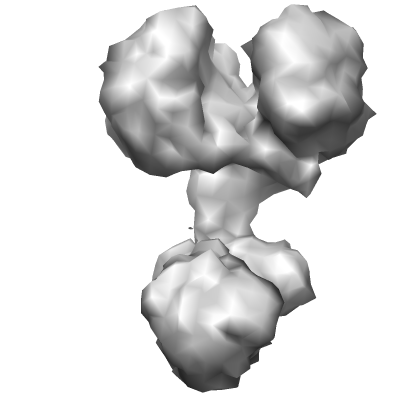 EMD-1985.png EMD-1985.png | 42.5 KB | ||
| Archive directory |  http://ftp.pdbj.org/pub/emdb/structures/EMD-1985ftp://ftp.pdbj.org/pub/emdb/structures/EMD-1985 http://ftp.pdbj.org/pub/emdb/structures/EMD-1985ftp://ftp.pdbj.org/pub/emdb/structures/EMD-1985 | HTTPS FTP |
-Related structure data
| Similar structure data |
|---|
-Links
| EMDB pages | EMDB (EBI/PDBe) / EMDataResource |
|---|
-Map
| File | Download / File: emd_1985.map.gz / Format: CCP4 / Size: 7.8 MB / Type: IMAGE STORED AS FLOATING POINT NUMBER (4 BYTES) | ||||||||||||||||||||||||||||||||||||||||||||||||||||||||||||||||||||
|---|---|---|---|---|---|---|---|---|---|---|---|---|---|---|---|---|---|---|---|---|---|---|---|---|---|---|---|---|---|---|---|---|---|---|---|---|---|---|---|---|---|---|---|---|---|---|---|---|---|---|---|---|---|---|---|---|---|---|---|---|---|---|---|---|---|---|---|---|---|
| Annotation | VDR-RXR DR3 DNA complex | ||||||||||||||||||||||||||||||||||||||||||||||||||||||||||||||||||||
| Voxel size | X=Y=Z: 2 Å | ||||||||||||||||||||||||||||||||||||||||||||||||||||||||||||||||||||
| Density |
| ||||||||||||||||||||||||||||||||||||||||||||||||||||||||||||||||||||
| Symmetry | Space group: 1 | ||||||||||||||||||||||||||||||||||||||||||||||||||||||||||||||||||||
| Details | EMDB XML:
CCP4 map header:
| ||||||||||||||||||||||||||||||||||||||||||||||||||||||||||||||||||||
-Supplemental data
- Sample components
-Entire : full human RXR-VDR nuclear receptor heterodimer complex with its ...
| Entire | Name: full human RXR-VDR nuclear receptor heterodimer complex with its DR3 target DNA response element |
|---|---|
| Components |
|
-Supramolecule #1000: full human RXR-VDR nuclear receptor heterodimer complex with its ...
| Supramolecule | Name: full human RXR-VDR nuclear receptor heterodimer complex with its DR3 target DNA response element type: sample / ID: 1000 / Number unique components: 3 |
|---|---|
| Molecular weight | Experimental: 100 KDa / Theoretical: 100 KDa |
-Macromolecule #1: VDR-RXR
| Macromolecule | Name: VDR-RXR / type: protein_or_peptide / ID: 1 / Name.synonym: VDR-RXR / Number of copies: 1 / Oligomeric state: hetero dimer / Recombinant expression: Yes |
|---|---|
| Source (natural) | Organism: Homo sapiens (human) / synonym: Human |
| Recombinant expression | Organism:  Escherichia coli (E. coli) / Recombinant plasmid: pACYC Escherichia coli (E. coli) / Recombinant plasmid: pACYC |
-Experimental details
-Structure determination
| Method | negative staining, cryo EM |
|---|---|
 Processing Processing | single particle reconstruction |
| Aggregation state | particle |
-Sample preparation
| Concentration | 0.15 mg/mL |
|---|---|
| Buffer | pH: 7.5 Details: Tris 20 mM pH7.5, NaCl 50 mM, KCl 50 mM, MgCl2 4mM, DTT 5mM |
| Staining | Type: NEGATIVE / Details: no staining, cryo on holey carbon film |
| Grid | Details: 300 mesh Cu/Rh |
| Vitrification | Cryogen name: ETHANE / Chamber humidity: 100 % / Instrument: OTHER / Details: Vitrification instrument: Vitrobot / Method: 2 seconds |
- Electron microscopy
| Microscope | FEI TECNAI F20 |
|---|---|
| Electron beam | Acceleration voltage: 200 kV / Electron source: FIELD EMISSION GUN |
| Electron optics | Illumination mode: FLOOD BEAM / Imaging mode: BRIGHT FIELDBright-field microscopy / Cs: 2.0 mm / Nominal defocus max: 4.0 µm / Nominal defocus min: 2.0 µm / Nominal magnification: 50000 |
| Sample stage | Specimen holder: Eucentric / Specimen holder model: GATAN LIQUID NITROGEN |
| Image recording | Category: FILM / Film or detector model: KODAK SO-163 FILM / Digitization - Scanner: PRIMESCAN / Digitization - Sampling interval: 5.8 µm / Number real images: 20 / Average electron dose: 20 e/Å2 / Bits/pixel: 16 |
| Experimental equipment |  Model: Tecnai F20 / Image courtesy: FEI Company |
-Image processing
| CTF correction | Details: each particle |
|---|---|
| Final reconstruction | Applied symmetry - Point group: C1 (asymmetric) / Algorithm: OTHER / Resolution.type: BY AUTHOR / Resolution: 12.0 Å / Resolution method: FSC 0.5 CUT-OFF / Software - Name: IMAGIC Details: resolution 12.3A or 9.1 according to FSC at 0.5 or 0.143 cut-off Number images used: 19938 |
| Details | EMAN-1 boxer semi-automatic selection and visual control of each boxed particle |
-Atomic model buiding 1
| Initial model | PDB ID: Chain - Chain ID: A |
|---|---|
| Software | Name: IMAGIC, pyMOL |
| Details | PDBEntryID_givenInChain. Protocol: rigid body. The domains were separately fitted by manual docking using the program Pymol |
| Refinement | Space: REAL / Protocol: RIGID BODY FIT |
-Atomic model buiding 2
| Initial model | PDB ID: Chain - Chain ID: A |
|---|---|
| Software | Name: IMAGIC, pyMOL |
| Details | PDBEntryID_givenInChain. Protocol: rigid body. The domains were separately fitted by manual docking using the program Pymol |
| Refinement | Space: REAL / Protocol: RIGID BODY FIT |
-Atomic model buiding 3
| Initial model | PDB ID: Chain - Chain ID: A |
|---|---|
| Software | Name: IMAGIC, pyMOL |
| Details | PDBEntryID_givenInChain. Protocol: rigid body. The domains were separately fitted by manual docking using the program Pymol |
| Refinement | Space: REAL / Protocol: RIGID BODY FIT |
-Atomic model buiding 4
| Initial model | PDB ID: Chain - Chain ID: A |
|---|---|
| Software | Name: IMAGIC, pyMOL |
| Details | PDBEntryID_givenInChain. Protocol: rigid body. The domains were separately fitted by manual docking using the program Pymol |
| Refinement | Space: REAL / Protocol: RIGID BODY FIT |