 Movie
Movie Controller


+ Open data

- Basic information
| Entry | Database: EMDB / ID: EMD-1913 | |||||||||
|---|---|---|---|---|---|---|---|---|---|---|
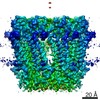
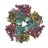
| Title | Structure of the Listeria monocytogenes ClpP1P2 complex | |||||||||
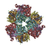
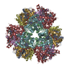
 Map data Map data | This is a surface rendered side view of ClpP1P2 | |||||||||
 Sample Sample |
| |||||||||
 Keywords Keywords | Proteomics / Vibralactone / heat shock protein / peptidase ClpP | |||||||||
| Biological species |  Listeria monocytogenes (bacteria) Listeria monocytogenes (bacteria) | |||||||||
| Method | single particle reconstruction / negative staining / Resolution: 15.0 Å | |||||||||
 Authors Authors | Zeiler E / Braun N / Boettcher T / Kastenmueller A / Weinkauf S / Sieber S | |||||||||
 Citation Citation | Journal: Angew Chem Int Ed Engl / Year: 2011 Title: Vibralactone as a tool to study the activity and structure of the ClpP1P2 complex from Listeria monocytogenes. Authors: Evelyn Zeiler / Nathalie Braun / Thomas Böttcher / Andreas Kastenmüller / Sevil Weinkauf / Stephan A Sieber /  | |||||||||
| History |
|
- Structure visualization
| Movie |
 Movie viewer Movie viewer |
|---|---|
| Structure viewer | EM map: SurfViewMolmilJmol/JSmol |
| Supplemental images |
 UCSF Chimera
- Downloads & links
-EMDB archive
| Map data | emd_1913.map.gz | 2.7 MB | EMDB map data format | |
|---|---|---|---|---|
| Header (meta data) | emd-1913-v30.xmlemd-1913.xml | 8.9 KB 8.9 KB | Display Display | EMDB header |
| Images | 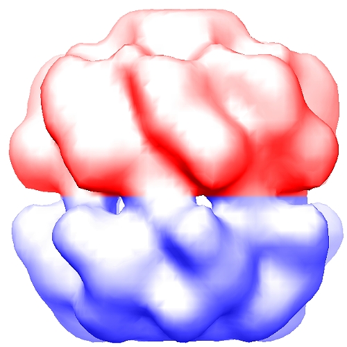 1913.jpg 1913.jpg | 126.6 KB | ||
| Archive directory |  http://ftp.pdbj.org/pub/emdb/structures/EMD-1913ftp://ftp.pdbj.org/pub/emdb/structures/EMD-1913 http://ftp.pdbj.org/pub/emdb/structures/EMD-1913ftp://ftp.pdbj.org/pub/emdb/structures/EMD-1913 | HTTPS FTP |
-Validation report
| Summary document | emd_1913_validation.pdf.gz | 202.7 KB | Display | EMDB validaton report |
|---|---|---|---|---|
| Full document | emd_1913_full_validation.pdf.gz | 201.8 KB | Display | |
| Data in XML | emd_1913_validation.xml.gz | 5.5 KB | Display | |
| Arichive directory | https://ftp.pdbj.org/pub/emdb/validation_reports/EMD-1913ftp://ftp.pdbj.org/pub/emdb/validation_reports/EMD-1913 | HTTPS FTP |
-Related structure data
| Similar structure data |
|---|

-Links
| EMDB pages | EMDB (EBI/PDBe) / EMDataResource |
|---|
-Map
| File | Download / File: emd_1913.map.gz / Format: CCP4 / Size: 7.8 MB / Type: IMAGE STORED AS FLOATING POINT NUMBER (4 BYTES) | ||||||||||||||||||||||||||||||||||||||||||||||||||||||||||||||||||||
|---|---|---|---|---|---|---|---|---|---|---|---|---|---|---|---|---|---|---|---|---|---|---|---|---|---|---|---|---|---|---|---|---|---|---|---|---|---|---|---|---|---|---|---|---|---|---|---|---|---|---|---|---|---|---|---|---|---|---|---|---|---|---|---|---|---|---|---|---|---|
| Annotation | This is a surface rendered side view of ClpP1P2 | ||||||||||||||||||||||||||||||||||||||||||||||||||||||||||||||||||||
| Voxel size | X=Y=Z: 1.46 Å | ||||||||||||||||||||||||||||||||||||||||||||||||||||||||||||||||||||
| Density |
| ||||||||||||||||||||||||||||||||||||||||||||||||||||||||||||||||||||
| Symmetry | Space group: 1 | ||||||||||||||||||||||||||||||||||||||||||||||||||||||||||||||||||||
| Details | EMDB XML:
CCP4 map header:
| ||||||||||||||||||||||||||||||||||||||||||||||||||||||||||||||||||||
-Supplemental data
- Sample components
-Entire : Listeria monocytogenes ClpP1P2 complex
| Entire | Name: Listeria monocytogenes ClpP1P2 complex |
|---|---|
| Components |
|
-Supramolecule #1000: Listeria monocytogenes ClpP1P2 complex
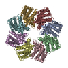
| Supramolecule | Name: Listeria monocytogenes ClpP1P2 complex / type: sample / ID: 1000 / Oligomeric state: Tetradecamer / Number unique components: 2 |
|---|---|
| Molecular weight | Experimental: 310 KDa / Theoretical: 310 KDa |
-Macromolecule #1: ClpP1
| Macromolecule | Name: ClpP1 / type: protein_or_peptide / ID: 1 / Name.synonym: ClpP1 / Number of copies: 1 / Oligomeric state: Heptamer / Recombinant expression: Yes |
|---|---|
| Source (natural) | Organism: Listeria monocytogenes (bacteria) |
-Macromolecule #2: ClpP2
| Macromolecule | Name: ClpP2 / type: protein_or_peptide / ID: 2 / Name.synonym: ClpP2 / Number of copies: 1 / Oligomeric state: Heptamer / Recombinant expression: Yes |
|---|---|
| Source (natural) | Organism: Listeria monocytogenes (bacteria) |
-Experimental details
-Structure determination
| Method | negative staining |
|---|---|
 Processing Processing | single particle reconstruction |
| Aggregation state | particle |
-Sample preparation
| Concentration | 0.2 mg/mL |
|---|---|
| Buffer | pH: 7 / Details: 50 mM MES, 100 mM KCl, 5% glycerol |
| Staining | Type: NEGATIVE Details: Grids with adsorbed protein floated on 1.5% w/v uranyl acetate for 30 seconds. |
| Vitrification | Cryogen name: NONE / Instrument: OTHER |
- Electron microscopy
| Microscope | JEOL 100CX |
|---|---|
| Alignment procedure | Legacy - Astigmatism: objective lens astigmatism was corrected at 100,000 times magnification |
| Image recording | Digitization - Scanner: OTHER / Number real images: 16 / Bits/pixel: 16 |
| Electron beam | Acceleration voltage: 100 kV / Electron source: TUNGSTEN HAIRPIN |
| Electron optics | Calibrated magnification: 58000 / Illumination mode: SPOT SCAN / Imaging mode: BRIGHT FIELD / Cs: 2.8 mm / Nominal defocus max: 1.0 µm / Nominal defocus min: 0.3 µm / Nominal magnification: 50000 |
| Sample stage | Specimen holder: eucentric / Specimen holder model: JEOL |
-Image processing
| CTF correction | Details: Phase flipping |
|---|---|
| Final reconstruction | Applied symmetry - Point group: C7 (7 fold cyclic) / Resolution.type: BY AUTHOR / Resolution: 15.0 Å / Resolution method: FSC 0.5 CUT-OFF / Software - Name: Imagic / Number images used: 39116 |
| Final two d classification | Number classes: 64 |
