 Movie
Movie Controller

[English] 日本語
 Yorodumi
- EMDB-1907: Electron cryo-microscopy and image reconstruction of adeno-associ... -
+ Open data

- Basic information
| Entry | Database: EMDB / ID: EMD-1907 | |||||||||
|---|---|---|---|---|---|---|---|---|---|---|
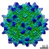
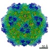
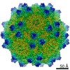
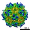
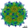
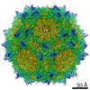
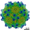
| Title | Electron cryo-microscopy and image reconstruction of adeno-associated virus type 2 empty capsids | |||||||||
 Map data Map data | AAV-2 empty capsids | |||||||||
 Sample Sample |
| |||||||||
 Keywords Keywords |  AAV2 / virus / empty capsids AAV2 / virus / empty capsids | |||||||||
| Biological species |  Adeno-associated virus - 2 Adeno-associated virus - 2 | |||||||||
| Method | single particle reconstruction / cryo EM / Resolution: 10.5 Å | |||||||||
 Authors Authors | Kronenberg S / Kleinschmidt JA / Bottcher B | |||||||||
 Citation Citation | Journal: EMBO Rep / Year: 2001 Title: Electron cryo-microscopy and image reconstruction of adeno-associated virus type 2 empty capsids. Authors: S Kronenberg / J A Kleinschmidt / B Böttcher /  Abstract: Adeno-associated virus type 2 empty capsids are composed of three proteins, VP1, VP2 and VP3, which have relative molecular masses of 87, 72 and 62 kDa, respectively, and differ in their N-terminal ...Adeno-associated virus type 2 empty capsids are composed of three proteins, VP1, VP2 and VP3, which have relative molecular masses of 87, 72 and 62 kDa, respectively, and differ in their N-terminal amino acid sequences. They have a likely molar ratio of 1:1:8 and occupy symmetrical equivalent positions in an icosahedrally arranged protein shell. We have investigated empty capsids of adeno-associated virus type 2 by electron cryo-microscopy and icosahedral image reconstruction. The three-dimensional map at 1.05 nm resolution showed sets of three elongated spikes surrounding the three-fold symmetry axes and narrow empty channels at the five-fold axes. The inside of the capsid superimposed with the previously determined structure of the canine parvovirus (Q. Xie and M.S. Chapman, 1996, J. Mol. Biol., 264, 497-520), whereas the outer surface showed clear discrepancies. Globular structures at the inner surface of the capsid at the two-fold symmetry axes were identified as possible positions for the N-terminal extensions of VP1 and VP2. | |||||||||
| History |
|
- Structure visualization
| Movie |
 Movie viewer Movie viewer |
|---|---|
| Structure viewer | EM map: SurfViewMolmilJmol/JSmol |
| Supplemental images |

- Downloads & links
-EMDB archive
| Map data | emd_1907.map.gz | 2 MB | EMDB map data format | |
|---|---|---|---|---|
| Header (meta data) | emd-1907-v30.xmlemd-1907.xml | 9.1 KB 9.1 KB | Display Display | EMDB header |
| Images | 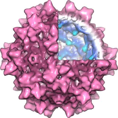 1907_aav2_1.jpg 1907_aav2_1.jpg | 26.9 KB | ||
| Archive directory |  http://ftp.pdbj.org/pub/emdb/structures/EMD-1907ftp://ftp.pdbj.org/pub/emdb/structures/EMD-1907 http://ftp.pdbj.org/pub/emdb/structures/EMD-1907ftp://ftp.pdbj.org/pub/emdb/structures/EMD-1907 | HTTPS FTP |
-Related structure data
| Similar structure data |
|---|
-Links
| EMDB pages | EMDB (EBI/PDBe) / EMDataResource |
|---|
-Map
| File | Download / File: emd_1907.map.gz / Format: CCP4 / Size: 2.1 MB / Type: IMAGE STORED AS FLOATING POINT NUMBER (4 BYTES) | ||||||||||||||||||||||||||||||||||||||||||||||||||||||||||||||||||||
|---|---|---|---|---|---|---|---|---|---|---|---|---|---|---|---|---|---|---|---|---|---|---|---|---|---|---|---|---|---|---|---|---|---|---|---|---|---|---|---|---|---|---|---|---|---|---|---|---|---|---|---|---|---|---|---|---|---|---|---|---|---|---|---|---|---|---|---|---|---|
| Annotation | AAV-2 empty capsids | ||||||||||||||||||||||||||||||||||||||||||||||||||||||||||||||||||||
| Voxel size | X=Y=Z: 4.2 Å | ||||||||||||||||||||||||||||||||||||||||||||||||||||||||||||||||||||
| Density |
| ||||||||||||||||||||||||||||||||||||||||||||||||||||||||||||||||||||
| Symmetry | Space group: 1 | ||||||||||||||||||||||||||||||||||||||||||||||||||||||||||||||||||||
| Details | EMDB XML:
CCP4 map header:
| ||||||||||||||||||||||||||||||||||||||||||||||||||||||||||||||||||||
-Supplemental data
- Sample components
-Entire : Adeno-associated Virus Type 2
| Entire | Name: Adeno-associated Virus Type 2 |
|---|---|
| Components |
|
-Supramolecule #1000: Adeno-associated Virus Type 2
| Supramolecule | Name: Adeno-associated Virus Type 2 / type: sample / ID: 1000 / Number unique components: 1 |
|---|---|
| Molecular weight | Theoretical: 3.9 MDa |
-Supramolecule #1: Adeno-associated virus - 2
| Supramolecule | Name: Adeno-associated virus - 2 / type: virus / ID: 1 / Name.synonym: AAV2 / NCBI-ID: 10804 / Sci species name: Adeno-associated virus - 2 / Virus type: VIRUS-LIKE PARTICLE / Virus isolate: SEROTYPE / Virus enveloped: No / Virus empty: Yes / Syn species name: AAV2 |
|---|---|
| Host (natural) | Organism:  Homo sapiens (human) / synonym: VERTEBRATES Homo sapiens (human) / synonym: VERTEBRATES |
| Molecular weight | Theoretical: 3.9 MDa |
| Virus shell | Shell ID: 1 / Name: AAV2 / Diameter: 260 Å / T number (triangulation number): 1 |
-Experimental details
-Structure determination
| Method | cryo EM |
|---|---|
 Processing Processing | single particle reconstruction |
| Aggregation state | particle |
-Sample preparation
| Buffer | pH: 7.5 / Details: 0.1M NaCl, 1 mM MgCl2, 10 mM Tris-HCl pH 7.5 |
|---|---|
| Grid | Details: 400 mesh copper grid, coated with holey carbon, covered with thin continuous carbon |
| Vitrification | Cryogen name: ETHANE / Chamber humidity: 100 % / Chamber temperature: 77 K / Instrument: HOMEMADE PLUNGER / Details: Vitrification instrument: Controlled environment / Method: blot for 15s before plunging |
- Electron microscopy
| Microscope | FEI/PHILIPS CM120T |
|---|---|
| Electron beam | Acceleration voltage: 100 kV / Electron source: LAB6 |
| Electron optics | Illumination mode: FLOOD BEAM / Imaging mode: BRIGHT FIELDBright-field microscopy / Cs: 6.4 mm / Nominal defocus max: 1.93 µm / Nominal defocus min: 0.81 µm / Nominal magnification: 52000 |
| Sample stage | Specimen holder: Side entry, liquid nitrogen cooled / Specimen holder model: GATAN LIQUID NITROGEN |
| Temperature | Min: 94 K / Average: 94 K |
| Alignment procedure | Legacy - Astigmatism: At 200,000 magnification on carbon |
| Date | Apr 10, 2001 |
| Image recording | Category: FILM / Film or detector model: KODAK SO-163 FILM / Digitization - Scanner: ZEISS SCAI / Digitization - Sampling interval: 21 µm / Number real images: 10 / Bits/pixel: 8 |
| Tilt angle min | 0 |
| Tilt angle max | 0 |
-Image processing
| CTF correction | Details: Combination of defocussed maps |
|---|---|
| Final reconstruction | Applied symmetry - Point group: I (icosahedral) / Algorithm: OTHER / Resolution.type: BY AUTHOR / Resolution: 10.5 Å / Resolution method: FSC 0.5 CUT-OFF / Software - Name: MRC Details: Maps were calculated for each micrograph maps were ctf-corrected and averaged ctf-weighted data was corrected for envelope function due to spatial aberration Number images used: 1800 |
