Movie
Movie Controller

[English] 日本語
 Yorodumi
- EMDB-1796: Proteomic characterization of archaeal ribosomes reveals the pres... -
+ Open data

- Basic information
| Entry | Database: EMDB / ID: EMD-1796 | |||||||||
|---|---|---|---|---|---|---|---|---|---|---|
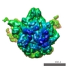
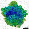
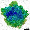
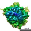
| Title | Proteomic characterization of archaeal ribosomes reveals the presence of novel archaeal-specific ribosomal proteins | |||||||||
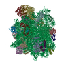
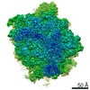
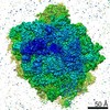
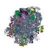
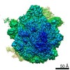
 Map data Map data | This is a 50S subunit of P.aerophilum Ribosome | |||||||||
 Sample Sample |
| |||||||||
 Keywords Keywords |  archaea / ribosome / 50S / translation / ribosomal proteins archaea / ribosome / 50S / translation / ribosomal proteins | |||||||||
| Biological species |   Pyrobaculum aerophilum (archaea) Pyrobaculum aerophilum (archaea) | |||||||||
| Method | single particle reconstruction / cryo EM / negative staining / Resolution: 25.0 Å | |||||||||
 Authors Authors | Marquez V / Froehlich T / Armache J-P / Sohmen D / Doenhoefer A / Berninghausen O / Thomm M / Beckmann R / Arnold GJ / Wilson DN | |||||||||
 Citation Citation | Journal: J Mol Biol / Year: 2011 Title: Proteomic characterization of archaeal ribosomes reveals the presence of novel archaeal-specific ribosomal proteins. Authors: Viter Márquez / Thomas Fröhlich / Jean-Paul Armache / Daniel Sohmen / Alexandra Dönhöfer / Aleksandra Mikolajka / Otto Berninghausen / Michael Thomm / Roland Beckmann / Georg J Arnold / Daniel N Wilson /  Abstract: Protein synthesis occurs in macromolecular particles called ribosomes. All ribosomes are composed of RNA and proteins. While the protein composition of bacterial and eukaryotic ribosomes has been ...Protein synthesis occurs in macromolecular particles called ribosomes. All ribosomes are composed of RNA and proteins. While the protein composition of bacterial and eukaryotic ribosomes has been well-characterized, a systematic analysis of archaeal ribosomes has been lacking. Here we report the first comprehensive two-dimensional PAGE and mass spectrometry analysis of archaeal ribosomes isolated from the thermophilic Pyrobaculum aerophilum and the thermoacidophilic Sulfolobus acidocaldarius Crenarchaeota. Our analysis identified all 66 ribosomal proteins (r-proteins) of the P. aerophilum small and large subunits, as well as all but two (62 of 64; 97%) r-proteins of the S. acidocaldarius small and large subunits that are predicted genomically. Some r-proteins were identified with one or two lysine methylations and N-terminal acetylations. In addition, we identify three hypothetical proteins that appear to be bona fide r-proteins of the S. acidocaldarius large subunit. Dissociation of r-proteins from the S. acidocaldarius large subunit indicates that the novel r-proteins establish tighter interactions with the large subunit than some integral r-proteins. Furthermore, cryo electron microscopy reconstructions of the S. acidocaldarius and P. aerophilum 50S subunits allow for a tentative localization of the binding site of the novel r-proteins. This study illustrates not only the potential diversity of the archaeal ribosomes but also the necessity to experimentally analyze the archaeal ribosomes to ascertain their protein composition. The discovery of novel archaeal r-proteins and factors may be the first step to understanding how archaeal ribosomes cope with extreme environmental conditions. | |||||||||
| History |
|
- Structure visualization
| Movie |
 Movie viewer Movie viewer |
|---|---|
| Structure viewer | EM map: SurfViewMolmilJmol/JSmol |
| Supplemental images |

- Downloads & links
-EMDB archive
| Map data | emd_1796.map.gz | 881.1 KB | EMDB map data format | |
|---|---|---|---|---|
| Header (meta data) | emd-1796-v30.xmlemd-1796.xml | 9.7 KB 9.7 KB | Display Display | EMDB header |
| Images | 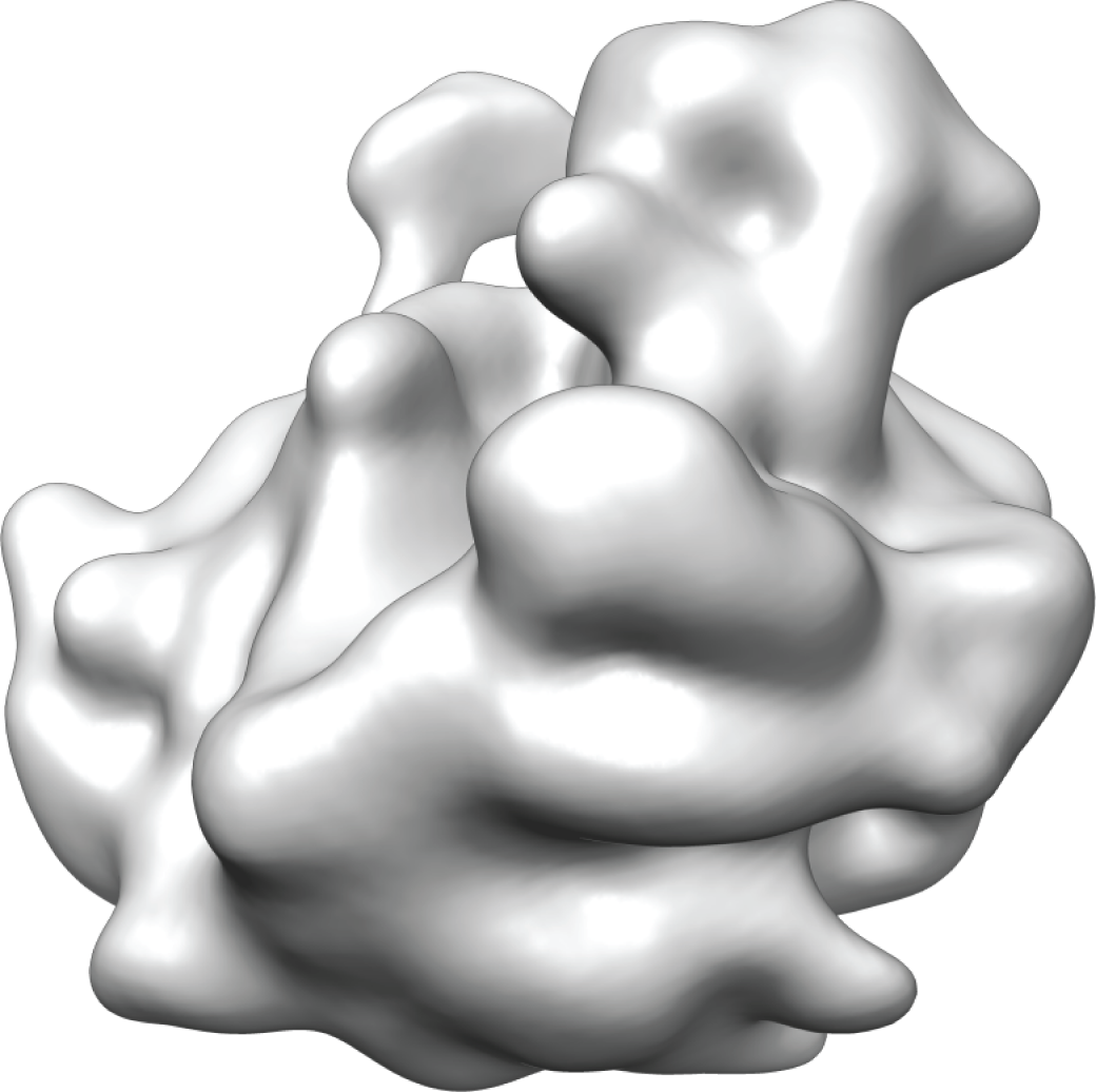 emd1796.png emd1796.png | 470.1 KB | ||
| Archive directory |  http://ftp.pdbj.org/pub/emdb/structures/EMD-1796ftp://ftp.pdbj.org/pub/emdb/structures/EMD-1796 http://ftp.pdbj.org/pub/emdb/structures/EMD-1796ftp://ftp.pdbj.org/pub/emdb/structures/EMD-1796 | HTTPS FTP |
-Related structure data

-Links
| EMDB pages | EMDB (EBI/PDBe) / EMDataResource |
|---|---|
| Related items in Molecule of the Month |

-Map
| File | Download / File: emd_1796.map.gz / Format: CCP4 / Size: 9.8 MB / Type: IMAGE STORED AS FLOATING POINT NUMBER (4 BYTES) | ||||||||||||||||||||||||||||||||||||||||||||||||||||||||||||||||||||
|---|---|---|---|---|---|---|---|---|---|---|---|---|---|---|---|---|---|---|---|---|---|---|---|---|---|---|---|---|---|---|---|---|---|---|---|---|---|---|---|---|---|---|---|---|---|---|---|---|---|---|---|---|---|---|---|---|---|---|---|---|---|---|---|---|---|---|---|---|---|
| Annotation | This is a 50S subunit of P.aerophilum Ribosome | ||||||||||||||||||||||||||||||||||||||||||||||||||||||||||||||||||||
| Voxel size | X=Y=Z: 3.308 Å | ||||||||||||||||||||||||||||||||||||||||||||||||||||||||||||||||||||
| Density |
| ||||||||||||||||||||||||||||||||||||||||||||||||||||||||||||||||||||
| Symmetry | Space group: 1 | ||||||||||||||||||||||||||||||||||||||||||||||||||||||||||||||||||||
| Details | EMDB XML:
CCP4 map header:
| ||||||||||||||||||||||||||||||||||||||||||||||||||||||||||||||||||||
-Supplemental data
- Sample components
-Entire : Pyrobaculum aerophilum 50S ribosomal subunit
| Entire | Name: Pyrobaculum aerophilum 50S ribosomal subunit |
|---|---|
| Components |
|
-Supramolecule #1000: Pyrobaculum aerophilum 50S ribosomal subunit
| Supramolecule | Name: Pyrobaculum aerophilum 50S ribosomal subunit / type: sample / ID: 1000 / Oligomeric state: One ribosomal subunit / Number unique components: 1 |
|---|---|
| Molecular weight | Experimental: 1.5 MDa / Theoretical: 1.5 MDa / Method: Sedimentation |
-Supramolecule #1: Pyrobaculum aerophilum 50S ribosomal subunit
| Supramolecule | Name: Pyrobaculum aerophilum 50S ribosomal subunit / type: complex / ID: 1 / Name.synonym: 50S of P.aerophilum / Recombinant expression: No / Ribosome-details: ribosome-prokaryote: LSU 50S |
|---|---|
| Source (natural) | Organism: Pyrobaculum aerophilum (archaea) |
| Molecular weight | Experimental: 1.5 MDa / Theoretical: 1.5 MDa |
-Experimental details
-Structure determination
| Method | negative staining, cryo EM |
|---|---|
 Processing Processing | single particle reconstruction |
| Aggregation state | particle |
-Sample preparation
| Buffer | pH: 7.5 Details: 20 mM Hepes pH 7.5, 10 mM Mg(OAc)2, 30 mM NH4OAc, 4 mM beta-Mercaptoethanol |
|---|---|
| Staining | Type: NEGATIVE / Details: Cryo-EM |
| Vitrification | Cryogen name: ETHANE / Chamber humidity: 100 % / Instrument: OTHER / Details: Vitrification instrument: Vitrobot Method: Blot for 10 seconds before plunging, use 2 layers of filter paper |
- Electron microscopy
| Microscope | FEI TECNAI 12 |
|---|---|
| Electron beam | Acceleration voltage: 120 kV / Electron source: LAB6 |
| Electron optics | Calibrated magnification: 90000 / Illumination mode: FLOOD BEAM / Imaging mode: BRIGHT FIELDBright-field microscopy / Cs: 2.0 mm / Nominal defocus max: 4.5 µm / Nominal defocus min: 1.0 µm / Nominal magnification: 90000 |
| Sample stage | Specimen holder: Single tilt cryo holder / Specimen holder model: GATAN LIQUID NITROGEN |
| Image recording | Category: CCD / Film or detector model: FEI EAGLE (2k x 2k) / Digitization - Sampling interval: 3.308 µm / Number real images: 99 / Average electron dose: 20 e/Å2 / Details: Data collected on CCD |
-Image processing
| CTF correction | Details: Wiener filter on 3D volumes |
|---|---|
| Final reconstruction | Applied symmetry - Point group: C1 (asymmetric) / Algorithm: OTHER / Resolution.type: BY AUTHOR / Resolution: 25.0 Å / Resolution method: FSC 0.5 CUT-OFF / Software - Name: SPIDER / Number images used: 9183 |
-Atomic model buiding 1
| Initial model | PDB ID: |
|---|---|
| Software | Name: UCSF Chimera |
| Details | Rigid-body fit into the density using UCSF Chimera built-in "Fit in Map" |
| Refinement | Space: REAL / Protocol: RIGID BODY FIT |