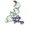
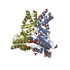
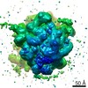
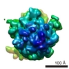
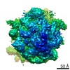
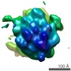
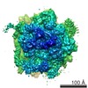
Journal: Nat Struct Mol Biol / Year: 2011 Title: Cryo-EM structure of the E. coli translating ribosome in complex with SRP and its receptor. Authors: Leandro F Estrozi / Daniel Boehringer / Shu-Ou Shan / Nenad Ban / Christiane Schaffitzel / Abstract: We report the 'early' conformation of the Escherichia coli signal recognition particle (SRP) and its receptor FtsY bound to the translating ribosome, as determined by cryo-EM. FtsY binds to the ...We report the 'early' conformation of the Escherichia coli signal recognition particle (SRP) and its receptor FtsY bound to the translating ribosome, as determined by cryo-EM. FtsY binds to the tetraloop of the SRP RNA, whereas the NG domains of the SRP protein and FtsY interact weakly in this conformation. Our results suggest that optimal positioning of the SRP RNA tetraloop and the Ffh NG domain leads to FtsY recruitment.
History
Deposition
Jul 13, 2010
-
Header (metadata) release
Nov 16, 2010
-
Map release
Mar 23, 2011
-
Update
Dec 11, 2013
-
Current status
Dec 11, 2013
Processing site: PDBe / Status: Released
-
Structure visualization
Movie
Surface view with section colored by density value
In the structure databanks used in Yorodumi, some data are registered as the other names, "COVID-19 virus" and "2019-nCoV". Here are the details of the virus and the list of structure data.
Jan 31, 2019. EMDB accession codes are about to change! (news from PDBe EMDB page)
EMDB accession codes are about to change! (news from PDBe EMDB page)
The allocation of 4 digits for EMDB accession codes will soon come to an end. Whilst these codes will remain in use, new EMDB accession codes will include an additional digit and will expand incrementally as the available range of codes is exhausted. The current 4-digit format prefixed with “EMD-” (i.e. EMD-XXXX) will advance to a 5-digit format (i.e. EMD-XXXXX), and so on. It is currently estimated that the 4-digit codes will be depleted around Spring 2019, at which point the 5-digit format will come into force.
The EM Navigator/Yorodumi systems omit the EMD- prefix.
Related info.:Q: What is EMD? / ID/Accession-code notation in Yorodumi/EM Navigator
Yorodumi is a browser for structure data from EMDB, PDB, SASBDB, etc.
This page is also the successor to EM Navigator detail page, and also detail information page/front-end page for Omokage search.
The word "yorodu" (or yorozu) is an old Japanese word meaning "ten thousand". "mi" (miru) is to see.
Related info.:EMDB / PDB / SASBDB / Comparison of 3 databanks / Yorodumi Search / Aug 31, 2016. New EM Navigator & Yorodumi / Yorodumi Papers / Jmol/JSmol / Function and homology information / Changes in new EM Navigator and Yorodumi
 Movie
Movie Controller

 Yorodumi
 Open data
 Basic information
 Map data
 Sample
 Keywords
 Ribosome / Co-translational Targeting /
  Function and homology information
 Authors
 Citation
 Structure visualization
 Downloads & links
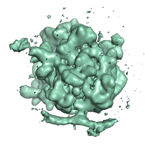 1762.png
 http://ftp.pdbj.org/pub/emdb/structures/EMD-1762


 Sample components
 Processing
 Electron microscopy