 Movie
Movie Controller

[English] 日本語
 Yorodumi
- EMDB-1712: Three-dimensional CryoEM Structure of a Membrane-Anchored AAA Protease -
+ Open data

- Basic information
| Entry | Database: EMDB / ID: EMD-1712 | ||||||||||||
|---|---|---|---|---|---|---|---|---|---|---|---|---|---|
| Title | Three-dimensional CryoEM Structure of a Membrane-Anchored AAA Protease | ||||||||||||
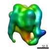
 Map data Map data | Isosurface representation of m-AAA protease. The density of transmembrane and intermembrane domains is weaker than those of AAA and protease domains. In the publication, the map was split into two parts and contoured separately, 91 kDa and 440 kDa. | ||||||||||||
 Sample Sample |
| ||||||||||||
 Keywords Keywords |  AAA protein / protease / ATPase / membrane protein / mitocondria AAA protein / protease / ATPase / membrane protein / mitocondria | ||||||||||||
| Biological species |  Saccharomyces cerevisiae (brewer's yeast) Saccharomyces cerevisiae (brewer's yeast) | ||||||||||||
| Method | single particle reconstruction / cryo EM / Resolution: 12.0 Å | ||||||||||||
 Authors Authors | Lee S / Augustin S / Tatsuta T / Gerdes F / Langer T / Tsai FTF | ||||||||||||
 Citation Citation | Journal: J Biol Chem / Year: 2011 Title: Electron cryomicroscopy structure of a membrane-anchored mitochondrial AAA protease. Authors: Sukyeong Lee / Steffen Augustin / Takashi Tatsuta / Florian Gerdes / Thomas Langer / Francis T F Tsai /  Abstract: FtsH-related AAA proteases are conserved membrane-anchored, ATP-dependent molecular machines, which mediate the processing and turnover of soluble and membrane-embedded proteins in eubacteria, ...FtsH-related AAA proteases are conserved membrane-anchored, ATP-dependent molecular machines, which mediate the processing and turnover of soluble and membrane-embedded proteins in eubacteria, mitochondria, and chloroplasts. Homo- and hetero-oligomeric proteolytic complexes exist, which are composed of homologous subunits harboring an ATPase domain of the AAA family and an H41 metallopeptidase domain. Mutations in subunits of mitochondrial m-AAA proteases have been associated with different neurodegenerative disorders in human, raising questions on the functional differences between homo- and hetero-oligomeric AAA proteases. Here, we have analyzed the hetero-oligomeric yeast m-AAA protease composed of homologous Yta10 and Yta12 subunits. We combined genetic and structural approaches to define the molecular determinants for oligomer assembly and to assess functional similarities between Yta10 and Yta12. We demonstrate that replacement of only two amino acid residues within the metallopeptidase domain of Yta12 allows its assembly into homo-oligomeric complexes. To provide a molecular explanation, we determined the 12 Å resolution structure of the intact yeast m-AAA protease with its transmembrane domains by electron cryomicroscopy (cryo-EM) and atomic structure fitting. The full-length m-AAA protease has a bipartite structure and is a hexamer in solution. We found that residues in Yta12, which facilitate homo-oligomerization when mutated, are located at the interface between neighboring protomers in the hexamer ring. Notably, the transmembrane and intermembrane space domains are separated from the main body, creating a passage on the matrix side, which is wide enough to accommodate unfolded but not folded polypeptides. These results suggest a mechanism regarding how proteins are recognized and degraded by m-AAA proteases. | ||||||||||||
| History |
|
- Structure visualization
| Movie |
 Movie viewer Movie viewer |
|---|---|
| Structure viewer | EM map: SurfViewMolmilJmol/JSmol |
| Supplemental images |

- Downloads & links
-EMDB archive
| Map data | emd_1712.map.gz | 1.7 MB | EMDB map data format | |
|---|---|---|---|---|
| Header (meta data) | emd-1712-v30.xmlemd-1712.xml | 10.9 KB 10.9 KB | Display Display | EMDB header |
| Images |  em1712.png em1712.png | 406.4 KB | ||
| Archive directory |  http://ftp.pdbj.org/pub/emdb/structures/EMD-1712ftp://ftp.pdbj.org/pub/emdb/structures/EMD-1712 http://ftp.pdbj.org/pub/emdb/structures/EMD-1712ftp://ftp.pdbj.org/pub/emdb/structures/EMD-1712 | HTTPS FTP |
-Related structure data
| Similar structure data |
|---|


-Links
| EMDB pages | EMDB (EBI/PDBe) / EMDataResource |
|---|
-Map
| File | Download / File: emd_1712.map.gz / Format: CCP4 / Size: 7.8 MB / Type: IMAGE STORED AS FLOATING POINT NUMBER (4 BYTES) | ||||||||||||||||||||||||||||||||||||||||||||||||||||||||||||||||||||
|---|---|---|---|---|---|---|---|---|---|---|---|---|---|---|---|---|---|---|---|---|---|---|---|---|---|---|---|---|---|---|---|---|---|---|---|---|---|---|---|---|---|---|---|---|---|---|---|---|---|---|---|---|---|---|---|---|---|---|---|---|---|---|---|---|---|---|---|---|---|
| Annotation | Isosurface representation of m-AAA protease. The density of transmembrane and intermembrane domains is weaker than those of AAA and protease domains. In the publication, the map was split into two parts and contoured separately, 91 kDa and 440 kDa. | ||||||||||||||||||||||||||||||||||||||||||||||||||||||||||||||||||||
| Voxel size | X=Y=Z: 1.81 Å | ||||||||||||||||||||||||||||||||||||||||||||||||||||||||||||||||||||
| Density |
| ||||||||||||||||||||||||||||||||||||||||||||||||||||||||||||||||||||
| Symmetry | Space group: 1 | ||||||||||||||||||||||||||||||||||||||||||||||||||||||||||||||||||||
| Details | EMDB XML:
CCP4 map header:
| ||||||||||||||||||||||||||||||||||||||||||||||||||||||||||||||||||||
-Supplemental data
- Sample components
-Entire : m-AAA Protease
| Entire | Name: m-AAA Protease |
|---|---|
| Components |
|
-Supramolecule #1000: m-AAA Protease
| Supramolecule | Name: m-AAA Protease / type: sample / ID: 1000 / Details: Sample was cross-linked / Oligomeric state: One hetero hexamer of Yta10 and Yta12 / Number unique components: 2 |
|---|---|
| Molecular weight | Experimental: 500 KDa / Theoretical: 500 KDa / Method: From the amino acid sequence |
-Macromolecule #1: Yta10
| Macromolecule | Name: Yta10 / type: protein_or_peptide / ID: 1 / Name.synonym: Yta10 / Details: Sample was cross-linked with Gluteraldehyde / Number of copies: 3 / Oligomeric state: hexamer / Recombinant expression: Yes |
|---|---|
| Source (natural) | Organism: Saccharomyces cerevisiae (brewer's yeast) / synonym: Baker's Yeast / Tissue: mitochondria / Cell: Saccharomyces cerevisiae / Organelle: mitochondria / Location in cell: mitochondrial inner membrane |
| Molecular weight | Experimental: 500 KDa / Theoretical: 500 KDa |
| Recombinant expression | Organism: Saccharomyces cerevisiae (brewer's yeast) |
-Macromolecule #2: Yta12
| Macromolecule | Name: Yta12 / type: protein_or_peptide / ID: 2 / Name.synonym: Yta12 / Details: Sample was cross-linked with Gluteraldehyde / Number of copies: 3 / Oligomeric state: hexamer / Recombinant expression: Yes |
|---|---|
| Source (natural) | Organism: Saccharomyces cerevisiae (brewer's yeast) / synonym: Baker's Yeast / Tissue: mitochondria / Cell: Saccharomyces cerevisiae / Organelle: mitochondria / Location in cell: mitochondrial inner membrane |
| Molecular weight | Experimental: 500 KDa / Theoretical: 500 KDa |
| Recombinant expression | Organism: Saccharomyces cerevisiae (brewer's yeast) |
-Experimental details
-Structure determination
| Method | cryo EM |
|---|---|
 Processing Processing | single particle reconstruction |
| Aggregation state | particle |
-Sample preparation
| Concentration | 0.3 mg/mL |
|---|---|
| Buffer | pH: 7.2 Details: 50mM Tris, pH 7.2, 150 mM NaCl, 1% n-octyl-beta-D-glucopyranoside, 10mM MgCl2, 5mM ATP |
| Grid | Details: 400 mesh cupper grid |
| Vitrification | Cryogen name: ETHANE / Chamber humidity: 100 % / Chamber temperature: 83 K / Instrument: OTHER / Details: Vitrification instrument: Vitrobot / Method: Blot for 5 seconds before plunging |
- Electron microscopy
| Microscope | JEOL 2010F |
|---|---|
| Electron beam | Acceleration voltage: 200 kV / Electron source: FIELD EMISSION GUN |
| Electron optics | Illumination mode: FLOOD BEAM / Imaging mode: BRIGHT FIELDBright-field microscopy / Nominal defocus max: 6.5 µm / Nominal defocus min: 1.8 µm / Nominal magnification: 60000 |
| Sample stage | Specimen holder: Eucentric / Specimen holder model: GATAN LIQUID NITROGEN |
| Temperature | Average: 93 K |
| Alignment procedure | Legacy - Astigmatism: objective lens astigmatism was corrected at 400,000 x magnification |
| Date | Jun 5, 2008 |
| Image recording | Category: CCD / Film or detector model: GENERIC GATAN (4k x 4k) / Average electron dose: 17 e/Å2 |
-Image processing
| CTF correction | Details: Each CCD image |
|---|---|
| Final two d classification | Number classes: 490 |
| Final reconstruction | Applied symmetry - Point group: C6 (6 fold cyclic) / Algorithm: OTHER / Resolution.type: BY AUTHOR / Resolution: 12.0 Å / Resolution method: OTHER / Software - Name: EMAN / Number images used: 37882 |
