Journal: Nature / Year: 2010 Title: Coupled chaperone action in folding and assembly of hexadecameric Rubisco. Authors: Cuimin Liu / Anna L Young / Amanda Starling-Windhof / Andreas Bracher / Sandra Saschenbrecker / Bharathi Vasudeva Rao / Karnam Vasudeva Rao / Otto Berninghausen / Thorsten Mielke / F Ulrich ...Authors: Cuimin Liu / Anna L Young / Amanda Starling-Windhof / Andreas Bracher / Sandra Saschenbrecker / Bharathi Vasudeva Rao / Karnam Vasudeva Rao / Otto Berninghausen / Thorsten Mielke / F Ulrich Hartl / Roland Beckmann / Manajit Hayer-Hartl / Abstract: Form I Rubisco (ribulose 1,5-bisphosphate carboxylase/oxygenase), a complex of eight large (RbcL) and eight small (RbcS) subunits, catalyses the fixation of atmospheric CO(2) in photosynthesis. The ...Form I Rubisco (ribulose 1,5-bisphosphate carboxylase/oxygenase), a complex of eight large (RbcL) and eight small (RbcS) subunits, catalyses the fixation of atmospheric CO(2) in photosynthesis. The limited catalytic efficiency of Rubisco has sparked extensive efforts to re-engineer the enzyme with the goal of enhancing agricultural productivity. To facilitate such efforts we analysed the formation of cyanobacterial form I Rubisco by in vitro reconstitution and cryo-electron microscopy. We show that RbcL subunit folding by the GroEL/GroES chaperonin is tightly coupled with assembly mediated by the chaperone RbcX(2). RbcL monomers remain partially unstable and retain high affinity for GroEL until captured by RbcX(2). As revealed by the structure of a RbcL(8)-(RbcX(2))(8) assembly intermediate, RbcX(2) acts as a molecular staple in stabilizing the RbcL subunits as dimers and facilitates RbcL(8) core assembly. Finally, addition of RbcS results in RbcX(2) release and holoenzyme formation. Specific assembly chaperones may be required more generally in the formation of complex oligomeric structures when folding is closely coupled to assembly.
History
Deposition
Oct 20, 2009
-
Header (metadata) release
Oct 27, 2009
-
Map release
Jan 20, 2010
-
Update
Jan 20, 2010
-
Current status
Jan 20, 2010
Processing site: PDBe / Status: Released
-
Structure visualization
Movie
Surface view with section colored by density value
Details: Quantifoil grids (3/3) with 2 nm carbon on top
Vitrification
Cryogen name: ETHANE / Chamber humidity: 95 % / Chamber temperature: 85 K / Instrument: OTHER / Details: Vitrification instrument: Vitrobot Method: 10 second blotting before plunging, used 2 layers of filter paper
-
Electron microscopy
Microscope
FEI TECNAI 12
Electron beam
Acceleration voltage: 120 kV / Electron source: LAB6
Category: CCD / Film or detector model: FEI EAGLE (2k x 2k) / Digitization - Sampling interval: 3.308 µm / Average electron dose: 20 e/Å2 / Details: Data collected on CCD / Bits/pixel: 16
Tilt angle max
0
-
Image processing
CTF correction
Details: Each particle
Final angle assignment
Details: EMAN
Final reconstruction
Applied symmetry - Point group: D4 (2x4 fold dihedral) / Algorithm: OTHER / Resolution.type: BY AUTHOR / Resolution: 16.0 Å / Resolution method: FSC 0.5 CUT-OFF / Software - Name: EMAN1 / Number images used: 13217
+
About Yorodumi
-
News
-
Feb 9, 2022. New format data for meta-information of EMDB entries
New format data for meta-information of EMDB entries
Version 3 of the EMDB header file is now the official format.
The previous official version 1.9 will be removed from the archive.
In the structure databanks used in Yorodumi, some data are registered as the other names, "COVID-19 virus" and "2019-nCoV". Here are the details of the virus and the list of structure data.
Jan 31, 2019. EMDB accession codes are about to change! (news from PDBe EMDB page)
EMDB accession codes are about to change! (news from PDBe EMDB page)
The allocation of 4 digits for EMDB accession codes will soon come to an end. Whilst these codes will remain in use, new EMDB accession codes will include an additional digit and will expand incrementally as the available range of codes is exhausted. The current 4-digit format prefixed with “EMD-” (i.e. EMD-XXXX) will advance to a 5-digit format (i.e. EMD-XXXXX), and so on. It is currently estimated that the 4-digit codes will be depleted around Spring 2019, at which point the 5-digit format will come into force.
The EM Navigator/Yorodumi systems omit the EMD- prefix.
Related info.:Q: What is EMD? / ID/Accession-code notation in Yorodumi/EM Navigator
Yorodumi is a browser for structure data from EMDB, PDB, SASBDB, etc.
This page is also the successor to EM Navigator detail page, and also detail information page/front-end page for Omokage search.
The word "yorodu" (or yorozu) is an old Japanese word meaning "ten thousand". "mi" (miru) is to see.
Related info.:EMDB / PDB / SASBDB / Comparison of 3 databanks / Yorodumi Search / Aug 31, 2016. New EM Navigator & Yorodumi / Yorodumi Papers / Jmol/JSmol / Function and homology information / Changes in new EM Navigator and Yorodumi
 Movie
Movie Controller


 Open data
 Basic information
 Map data
 Sample
 Keywords
 photosynthesis /
  Function and homology information
 Authors
 Citation
 Structure visualization
 Downloads & links
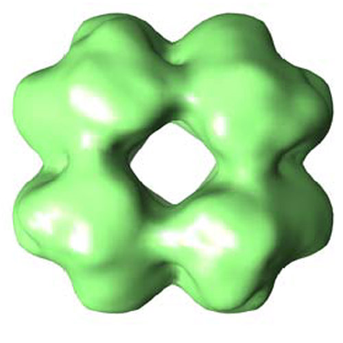 emd_1656.jpg
 http://ftp.pdbj.org/pub/emdb/structures/EMD-1656


 Z (Sec.)
 Y (Row.)
 X (Col.)


 Sample components
 Processing
 Electron microscopy