 ムービー
ムービー コントローラー


+ データを開く

- 基本情報
基本情報
| 登録情報 | データベース: EMDB / ID: EMD-1640 | |||||||||
|---|---|---|---|---|---|---|---|---|---|---|
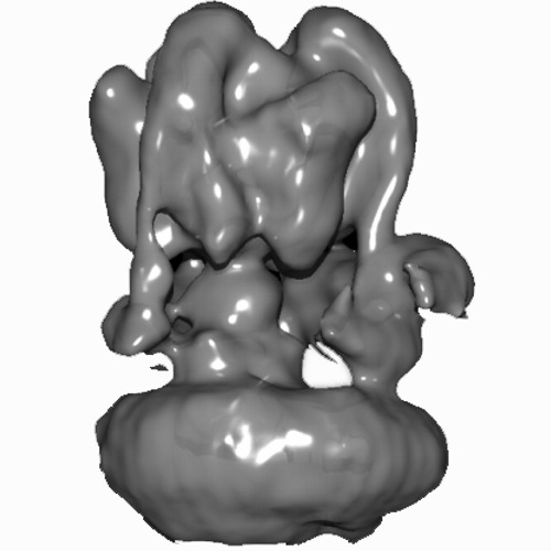
| タイトル | Structure of the V-ATPase of Saccharomyces cerevisiae at 2.5 nm resolution | |||||||||
 マップデータ マップデータ | V-ATPase of Sacharomyces cerevisiae | |||||||||
 試料 試料 |
| |||||||||
 キーワード キーワード | V-ATPase / single particle / Stator | |||||||||
| 生物種 |  | |||||||||
| 手法 | 単粒子再構成法 / ネガティブ染色法 / 解像度: 25.0 Å | |||||||||
 データ登録者 データ登録者 | Diepholz M / Venzke D / Prinz S / Batisse C / Florchinger B / Rossle M / Svergun D / Bottcher B / Fethiere J | |||||||||
 引用 引用 | ジャーナル: Structure / 年: 2008 タイトル: A different conformation for EGC stator subcomplex in solution and in the assembled yeast V-ATPase: possible implications for regulatory disassembly. 著者: Meikel Diepholz / David Venzke / Simone Prinz / Claire Batisse / Beate Flörchinger / Manfred Rössle / Dmitri I Svergun / Bettina Böttcher / James Féthière /  要旨: Vacuolar ATPases (V-ATPases) are ATP-dependent proton pumps that maintain the acidity of cellular compartments. They are composed of a membrane-integrated proton-translocating V(0) and an extrinsic ...Vacuolar ATPases (V-ATPases) are ATP-dependent proton pumps that maintain the acidity of cellular compartments. They are composed of a membrane-integrated proton-translocating V(0) and an extrinsic cytoplasmic catalytic domain V(1), joined by several connecting subunits. To clarify the arrangement of these peripheral connections and their interrelation with other subunits of the holocomplex, we have determined the solution structures of isolated EG and EGC connecting subcomplexes by small angle X-ray scattering and the 3D map of the yeast V-ATPase by electron microscopy. In solution, EG forms a slightly kinked rod, which assembles with subunit C into an L-shaped structure. This model is supported by the microscopy data, which show three copies of EG with two of these linked by subunit C. However, the relative arrangement of the EG and C subunits in solution is more open than that in the holoenzyme, suggesting a conformational change of EGC during regulatory assembly and disassembly. | |||||||||
| 履歴 |
|
- 構造の表示
構造の表示
| ムービー |
 ムービービューア ムービービューア |
|---|---|
| 構造ビューア | EMマップ: SurfViewMolmilJmol/JSmol |
| 添付画像 |
- ダウンロードとリンク
-EMDBアーカイブ
| マップデータ | emd_1640.map.gz | 271.6 KB | EMDBマップデータ形式 | |
|---|---|---|---|---|
| ヘッダ (付随情報) | emd-1640-v30.xmlemd-1640.xml | 9.7 KB 9.7 KB | 表示 表示 | EMDBヘッダ |
| 画像 | vatpase_emdb.tif | 137.4 KB | ||
| アーカイブディレクトリ |  http://ftp.pdbj.org/pub/emdb/structures/EMD-1640ftp://ftp.pdbj.org/pub/emdb/structures/EMD-1640 http://ftp.pdbj.org/pub/emdb/structures/EMD-1640ftp://ftp.pdbj.org/pub/emdb/structures/EMD-1640 | HTTPS FTP |
-検証レポート
| 文書・要旨 | emd_1640_validation.pdf.gz | 198.3 KB | 表示 | EMDB検証レポート |
|---|---|---|---|---|
| 文書・詳細版 | emd_1640_full_validation.pdf.gz | 197.4 KB | 表示 | |
| XML形式データ | emd_1640_validation.xml.gz | 5.2 KB | 表示 | |
| アーカイブディレクトリ | https://ftp.pdbj.org/pub/emdb/validation_reports/EMD-1640ftp://ftp.pdbj.org/pub/emdb/validation_reports/EMD-1640 | HTTPS FTP |
-関連構造データ


-リンク
| EMDBのページ | EMDB (EBI/PDBe) / EMDataResource |
|---|
-マップ
| ファイル | ダウンロード / ファイル: emd_1640.map.gz / 形式: CCP4 / 大きさ: 3.7 MB / タイプ: IMAGE STORED AS FLOATING POINT NUMBER (4 BYTES) | ||||||||||||||||||||||||||||||||||||||||||||||||||||||||||||||||||||
|---|---|---|---|---|---|---|---|---|---|---|---|---|---|---|---|---|---|---|---|---|---|---|---|---|---|---|---|---|---|---|---|---|---|---|---|---|---|---|---|---|---|---|---|---|---|---|---|---|---|---|---|---|---|---|---|---|---|---|---|---|---|---|---|---|---|---|---|---|---|
| 注釈 | V-ATPase of Sacharomyces cerevisiae | ||||||||||||||||||||||||||||||||||||||||||||||||||||||||||||||||||||
| ボクセルのサイズ | X=Y=Z: 5.17 Å | ||||||||||||||||||||||||||||||||||||||||||||||||||||||||||||||||||||
| 密度 |
| ||||||||||||||||||||||||||||||||||||||||||||||||||||||||||||||||||||
| 対称性 | 空間群: 1 | ||||||||||||||||||||||||||||||||||||||||||||||||||||||||||||||||||||
| 詳細 | EMDB XML:
CCP4マップ ヘッダ情報:
| ||||||||||||||||||||||||||||||||||||||||||||||||||||||||||||||||||||
-添付データ
- 試料の構成要素
-全体 : V-ATPase
| 全体 | 名称: V-ATPase |
|---|---|
| 要素 |
|
-超分子 #1000: V-ATPase
| 超分子 | 名称: V-ATPase / タイプ: sample / ID: 1000 / 集合状態: A3B3CDE3FG3Hac'c''c(5-8)d / Number unique components: 1 |
|---|---|
| 分子量 | 理論値: 980 KDa |
-分子 #1: V-ATPase
| 分子 | 名称: V-ATPase / タイプ: protein_or_peptide / ID: 1 / Name.synonym: V-ATPase / 組換発現: Yes |
|---|---|
| 由来(天然) | 生物種: |
| 組換発現 | 生物種: |
-実験情報
-構造解析
| 手法 | ネガティブ染色法 |
|---|---|
 解析 解析 | 単粒子再構成法 |
| 試料の集合状態 | particle |
-試料調製
| 緩衝液 | pH: 7.1 詳細: Phosphate buffered Saline containing 0.1% Digitonin, 8% sucrose, 2% sorbitol, 2% glucose, Pefabloc SC (Roche), 13 tablets/l of complete EDTA-free Protease inhibitors |
|---|---|
| 染色 | タイプ: NEGATIVE 詳細: The sandwich technique (Golas et al., 2003) was used to prepare negatively stained V-ATPase samples. Briefly, purified V-ATPase at a concentration of 0.03 mg/ml was adsorbed on a carbon film ...詳細: The sandwich technique (Golas et al., 2003) was used to prepare negatively stained V-ATPase samples. Briefly, purified V-ATPase at a concentration of 0.03 mg/ml was adsorbed on a carbon film layered on a mica support at the carbon/mica interface. Subsequently, the carbon film with adsorbed particles was floated over the staining solution (2% uranyl acetate). After attachment of a copper grid to the dry surface of the carbon, a second carbon film was floated over the same staining solution. The protein/carbon/grid assembly was picked up with a piece of newspaper, turned upside down, and immersed in the solution. A sandwich of two carbon layers with the protein particles trapped in between is created by picking up the second carbon layer with the grid. |
| グリッド | 詳細: 400 mesh copper grid |
| 凍結 | 凍結剤: NONE / 装置: OTHER |
- 電子顕微鏡法
| 顕微鏡 | FEI/PHILIPS CM200FEG |
|---|---|
| アライメント法 | Legacy - 非点収差: Corrected at 200000 times magnification on graininess of carbon |
| 撮影 | カテゴリ: CCD / フィルム・検出器のモデル: GENERIC CCD / デジタル化 - サンプリング間隔: 14.22 µm / 実像数: 400 詳細: Images were recorded on CCD, no scanning, sampling step size was adjusted to calibrated image size ビット/ピクセル: 12 |
| Tilt angle min | 0 |
| Tilt angle max | 0 |
| 電子線 | 加速電圧: 200 kV / 電子線源:  FIELD EMISSION GUN FIELD EMISSION GUN |
| 電子光学系 | 照射モード: FLOOD BEAM / 撮影モード: BRIGHT FIELD / Cs: 2 mm / 倍率(公称値): 27500 |
| 試料ステージ | 試料ホルダー: Eucentric / 試料ホルダーモデル: SIDE ENTRY, EUCENTRIC |
-画像解析
| 最終 再構成 | 想定した対称性 - 点群: C1 (非対称) / アルゴリズム: OTHER / 解像度のタイプ: BY AUTHOR / 解像度: 25.0 Å / 解像度の算出法: FSC 0.5 CUT-OFF / ソフトウェア - 名称: IMAGIC, SPIDER, EMAN 詳細: Spider option BP 32F Back Projection - 3D, Sampled, Interpolated in Fourier space 使用した粒子像数: 16300 |
|---|
