 Movie
Movie Controller

[English] 日本語
 Yorodumi
- EMDB-1605: Solution structure of the KdpFABC P-type ATPase from Escherichia ... -
+ Open data

- Basic information
| Entry | Database: EMDB / ID: EMD-1605 | |||||||||
|---|---|---|---|---|---|---|---|---|---|---|
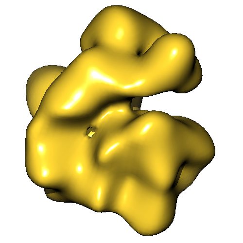
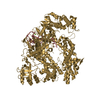
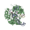
| Title | Solution structure of the KdpFABC P-type ATPase from Escherichia coli by electron microscopic single particle analysis | |||||||||
 Map data Map data | Volume of the KdpFABC P-type ATPase | |||||||||
 Sample Sample |
| |||||||||
 Keywords Keywords | KdpFABC /  P-type ATPase / potassium transport / single particle analysis / electron microscopy. P-type ATPase / potassium transport / single particle analysis / electron microscopy. | |||||||||
| Biological species |  Escherichia coli (E. coli) Escherichia coli (E. coli) | |||||||||
| Method | single particle reconstruction / negative staining / Resolution: 19.0 Å | |||||||||
 Authors Authors | Heitkamp T / Bottcher B / Greie J-C | |||||||||
 Citation Citation | Journal: J Struct Biol / Year: 2009 Title: Solution structure of the KdpFABC P-type ATPase from Escherichia coli by electron microscopic single particle analysis. Authors: Thomas Heitkamp / Bettina Böttcher / Jörg-Christian Greie /  Abstract: The K+-translocating KdpFABC complex from Escherichia coli functions as a high affinity potassium uptake system and belongs to the superfamily of P-type ATPases, although it exhibits some unique ...The K+-translocating KdpFABC complex from Escherichia coli functions as a high affinity potassium uptake system and belongs to the superfamily of P-type ATPases, although it exhibits some unique features. It comprises four subunits, and the sites of ATP hydrolysis and substrate transport are located on two different polypeptides. No structural data are so far available for elucidating the correspondingly unique mechanism of coupling ion transport and catalysis in this P-type ATPase. By use of electron microscopy and single particle analysis of negatively stained, solubilized KdpFABC complexes, we solved the structure of the complex at a resolution of 19A, which allowed us to model the arrangement of subunits within the holoenzyme and, thus, to identify the interfaces between subunits. The model showed that the K+-translocating KdpA subunit is in close contact with the transmembrane region of the ATP-hydrolyzing subunit KdpB. The cytosolic C-terminal domain of the KdpC subunit, which is assumed to play a role in cooperative ATP binding together with KdpB, is located in close vicinity to the nucleotide binding domain of KdpB. Overall, the arrangement of subunits agrees with biochemical data and the predictions on subunit interactions. | |||||||||
| History |
|
- Structure visualization
| Movie |
 Movie viewer Movie viewer |
|---|---|
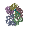
| Structure viewer | EM map: SurfViewMolmilJmol/JSmol |
| Supplemental images |
- Downloads & links
-EMDB archive
| Map data | emd_1605.map.gz | 167 KB | EMDB map data format | |
|---|---|---|---|---|
| Header (meta data) | emd-1605-v30.xmlemd-1605.xml | 11.9 KB 11.9 KB | Display Display | EMDB header |
| Images |  image.gif image.gif | 91.4 KB | ||
| Archive directory |  http://ftp.pdbj.org/pub/emdb/structures/EMD-1605ftp://ftp.pdbj.org/pub/emdb/structures/EMD-1605 http://ftp.pdbj.org/pub/emdb/structures/EMD-1605ftp://ftp.pdbj.org/pub/emdb/structures/EMD-1605 | HTTPS FTP |
-Related structure data
| Similar structure data |
|---|


-Links
| EMDB pages | EMDB (EBI/PDBe) / EMDataResource |
|---|
-Map
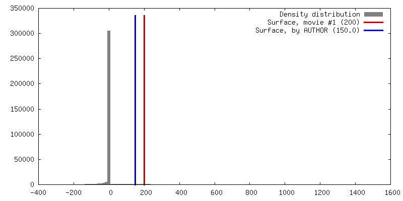
| File | Download / File: emd_1605.map.gz / Format: CCP4 / Size: 1.3 MB / Type: IMAGE STORED AS FLOATING POINT NUMBER (4 BYTES) | ||||||||||||||||||||||||||||||||||||||||||||||||||||||||||||||||||||
|---|---|---|---|---|---|---|---|---|---|---|---|---|---|---|---|---|---|---|---|---|---|---|---|---|---|---|---|---|---|---|---|---|---|---|---|---|---|---|---|---|---|---|---|---|---|---|---|---|---|---|---|---|---|---|---|---|---|---|---|---|---|---|---|---|---|---|---|---|---|
| Annotation | Volume of the KdpFABC P-type ATPase | ||||||||||||||||||||||||||||||||||||||||||||||||||||||||||||||||||||
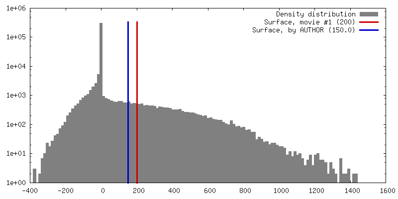
| Projections & slices | Image control
Images are generated by Spider. | ||||||||||||||||||||||||||||||||||||||||||||||||||||||||||||||||||||
| Voxel size | X=Y=Z: 2.8 Å | ||||||||||||||||||||||||||||||||||||||||||||||||||||||||||||||||||||
| Density |
| ||||||||||||||||||||||||||||||||||||||||||||||||||||||||||||||||||||
| Symmetry | Space group: 1 | ||||||||||||||||||||||||||||||||||||||||||||||||||||||||||||||||||||
| Details | EMDB XML:
CCP4 map header:
| ||||||||||||||||||||||||||||||||||||||||||||||||||||||||||||||||||||
 Z (Sec.)
 Y (Row.)
 X (Col.)


-Supplemental data
- Sample components
-Entire : KdpFABC P-type ATPase
| Entire | Name: KdpFABC P-type ATPase |
|---|---|
| Components |
|
-Supramolecule #1000: KdpFABC P-type ATPase
| Supramolecule | Name: KdpFABC P-type ATPase / type: sample / ID: 1000 / Oligomeric state: monomer / Number unique components: 1 |
|---|---|
| Molecular weight | Theoretical: 154 KDa |
-Macromolecule #1: KdpA-subunit
| Macromolecule | Name: KdpA-subunit / type: protein_or_peptide / ID: 1 / Name.synonym: KdpA-subunit / Number of copies: 1 / Oligomeric state: monomer / Recombinant expression: Yes |
|---|---|
| Source (natural) | Organism: Escherichia coli (E. coli) / Location in cell: membrane |
| Molecular weight | Theoretical: 59 KDa |
| Recombinant expression | Organism: Escherichia coli (E. coli) / Recombinant plasmid: pGS4 |
-Macromolecule #2: KdpB-subunit
| Macromolecule | Name: KdpB-subunit / type: protein_or_peptide / ID: 2 / Name.synonym: KdpB-subunit / Number of copies: 1 / Oligomeric state: monomer / Recombinant expression: Yes |
|---|---|
| Source (natural) | Organism: Escherichia coli (E. coli) / Location in cell: membrane |
| Molecular weight | Theoretical: 72 KDa |
| Recombinant expression | Organism: Escherichia coli (E. coli) / Recombinant plasmid: pGS4 |
-Macromolecule #3: KdpC-subunit
| Macromolecule | Name: KdpC-subunit / type: protein_or_peptide / ID: 3 / Name.synonym: KdpC-subunit / Number of copies: 1 / Oligomeric state: monomer / Recombinant expression: Yes |
|---|---|
| Source (natural) | Organism: Escherichia coli (E. coli) / Location in cell: membrane |
| Molecular weight | Theoretical: 21 KDa |
| Recombinant expression | Organism: Escherichia coli (E. coli) / Recombinant plasmid: pGS4 |
-Macromolecule #4: KdpF-subunit
| Macromolecule | Name: KdpF-subunit / type: protein_or_peptide / ID: 4 / Name.synonym: KdpF-subunit / Number of copies: 1 / Oligomeric state: monomer / Recombinant expression: Yes |
|---|---|
| Source (natural) | Organism: Escherichia coli (E. coli) / Location in cell: membrane |
| Molecular weight | Theoretical: 3 KDa |
| Recombinant expression | Organism: Escherichia coli (E. coli) / Recombinant plasmid: pGS4 |
-Experimental details
-Structure determination
| Method | negative staining |
|---|---|
 Processing Processing | single particle reconstruction |
| Aggregation state | particle |
-Sample preparation
| Concentration | 0.005 mg/mL |
|---|---|
| Buffer | pH: 6 Details: 50 mM MES pH 6.0, 50 mM NaCl, 5 mM MgCl2, 5 mM CaCl2 |
| Staining | Type: NEGATIVE Details: applied to freshly glow-discharged carbon-coated copper grids (400 mesh). Staining with 2 % (w/v) uranylacetic acid |
| Grid | Details: 400 mesh copper grid, carbon coated |
| Vitrification | Cryogen name: NONE / Instrument: OTHER |
- Electron microscopy
| Microscope | FEI/PHILIPS CM120T |
|---|---|
| Electron beam | Acceleration voltage: 100 kV / Electron source: LAB6 |
| Electron optics | Calibrated magnification: 48000 / Illumination mode: FLOOD BEAM / Imaging mode: BRIGHT FIELDBright-field microscopy / Cs: 2 mm / Nominal defocus max: 0.864 µm / Nominal defocus min: 0.432 µm / Nominal magnification: 52000 |
| Sample stage | Specimen holder: room temperature holder / Specimen holder model: SIDE ENTRY, EUCENTRIC |
| Temperature | Min: 95 K / Max: 295 K / Average: 295 K |
| Alignment procedure | Legacy - Astigmatism: manually at 200000 on carbon film |
| Date | Jul 20, 2004 |
| Image recording | Category: FILM / Film or detector model: KODAK SO-163 FILM / Digitization - Scanner: ZEISS SCAI / Digitization - Sampling interval: 14 µm / Number real images: 90 / Bits/pixel: 8 |
| Tilt angle min | 0 |
| Tilt angle max | 0 |
-Image processing
| Final reconstruction | Applied symmetry - Point group: C1 (asymmetric) / Algorithm: OTHER / Resolution.type: BY AUTHOR / Resolution: 19.0 Å / Resolution method: FSC 0.5 CUT-OFF / Software - Name: IMAGIC Spider / Number images used: 10040 |
|---|---|
| Details | started with angular reconstitution in IMAGIC (25 class averages) followed by projection matching in Spider (global search in 10 degree steps followed by local search in 2 degree steps) |
