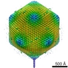
Journal: J Mol Biol / Year: 2009 Title: The capsid proteins of a large, icosahedral dsDNA virus. Authors: Xiaodong Yan / Zeyun Yu / Ping Zhang / Anthony J Battisti / Heather A Holdaway / Paul R Chipman / Chandrajit Bajaj / Max Bergoin / Michael G Rossmann / Timothy S Baker / Abstract: Chilo iridescent virus (CIV) is a large (approximately 1850 A diameter) insect virus with an icosahedral, T=147 capsid, a double-stranded DNA (dsDNA) genome, and an internal lipid membrane. The ...Chilo iridescent virus (CIV) is a large (approximately 1850 A diameter) insect virus with an icosahedral, T=147 capsid, a double-stranded DNA (dsDNA) genome, and an internal lipid membrane. The structure of CIV was determined to 13 A resolution by means of cryoelectron microscopy (cryoEM) and three-dimensional image reconstruction. A homology model of P50, the CIV major capsid protein (MCP), was built based on its amino acid sequence and the structure of the homologous Paramecium bursaria chlorella virus 1 Vp54 MCP. This model was fitted into the cryoEM density for each of the 25 trimeric CIV capsomers per icosahedral asymmetric unit. A difference map, in which the fitted CIV MCP capsomers were subtracted from the CIV cryoEM reconstruction, showed that there are at least three different types of minor capsid proteins associated with the capsomers outside the lipid membrane. "Finger" proteins are situated at many, but not all, of the spaces between three adjacent capsomers within each trisymmetron, and "zip" proteins are situated between sets of three adjacent capsomers at the boundary between neighboring trisymmetrons and pentasymmetrons. Based on the results of segmentation and density correlations, there are at least eight finger proteins and three dimeric and two monomeric zip proteins in one asymmetric unit of the CIV capsid. These minor proteins appear to stabilize the virus by acting as intercapsomer cross-links. One transmembrane "anchor" protein per icosahedral asymmetric unit, which extends from beneath one of the capsomers in the pentasymmetron to the internal leaflet of the lipid membrane, may provide additional stabilization for the capsid. These results are consistent with the observations for other large, icosahedral dsDNA viruses that also utilize minor capsid proteins for stabilization and for determining their assembly.
History
Deposition
Nov 4, 2008
-
Header (metadata) release
Nov 5, 2008
-
Map release
Apr 1, 2009
-
Update
Sep 9, 2011
-
Current status
Sep 9, 2011
Processing site: PDBe / Status: Released
-
Structure visualization
Movie
Surface view with section colored by density value
Category: FILM / Film or detector model: KODAK SO-163 FILM / Digitization - Scanner: ZEISS SCAI / Digitization - Sampling interval: 7.0 µm / Number real images: 210 / Average electron dose: 22 e/Å2 / Bits/pixel: 12
-
Image processing
CTF correction
Details: each micrograph
Final two d classification
Number classes: 1
Final reconstruction
Applied symmetry - Point group: I (icosahedral) / Algorithm: OTHER / Resolution.type: BY AUTHOR / Resolution: 13.0 Å / Resolution method: FSC 0.5 CUT-OFF / Software - Name: AUTO3DEM / Number images used: 1800
+
About Yorodumi
-
News
-
Feb 9, 2022. New format data for meta-information of EMDB entries
New format data for meta-information of EMDB entries
Version 3 of the EMDB header file is now the official format.
The previous official version 1.9 will be removed from the archive.
In the structure databanks used in Yorodumi, some data are registered as the other names, "COVID-19 virus" and "2019-nCoV". Here are the details of the virus and the list of structure data.
Jan 31, 2019. EMDB accession codes are about to change! (news from PDBe EMDB page)
EMDB accession codes are about to change! (news from PDBe EMDB page)
The allocation of 4 digits for EMDB accession codes will soon come to an end. Whilst these codes will remain in use, new EMDB accession codes will include an additional digit and will expand incrementally as the available range of codes is exhausted. The current 4-digit format prefixed with “EMD-” (i.e. EMD-XXXX) will advance to a 5-digit format (i.e. EMD-XXXXX), and so on. It is currently estimated that the 4-digit codes will be depleted around Spring 2019, at which point the 5-digit format will come into force.
The EM Navigator/Yorodumi systems omit the EMD- prefix.
Related info.:Q: What is EMD? / ID/Accession-code notation in Yorodumi/EM Navigator
Yorodumi is a browser for structure data from EMDB, PDB, SASBDB, etc.
This page is also the successor to EM Navigator detail page, and also detail information page/front-end page for Omokage search.
The word "yorodu" (or yorozu) is an old Japanese word meaning "ten thousand". "mi" (miru) is to see.
Related info.:EMDB / PDB / SASBDB / Comparison of 3 databanks / Yorodumi Search / Aug 31, 2016. New EM Navigator & Yorodumi / Yorodumi Papers / Jmol/JSmol / Function and homology information / Changes in new EM Navigator and Yorodumi
 Movie
Movie Controller

 Yorodumi
 Open data
 Basic information
 Map data
 Sample
 Keywords
 cryo-electron microscopy / 3D image reconstruction /
  Invertebrate iridescent virus 6
 Authors
 Citation
 Structure visualization
 Movie viewer
 Downloads & links
 1580.gif
 http://ftp.pdbj.org/pub/emdb/structures/EMD-1580
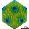
 Sample components
 Processing
 Electron microscopy