 Movie
Movie Controller

[English] 日本語
 Yorodumi
- EMDB-1550: Three-dimensional icosahedral reconstruction of Rift Valley Fever... -
+ Open data

- Basic information
| Entry | Database: EMDB / ID: EMD-1550 | |||||||||
|---|---|---|---|---|---|---|---|---|---|---|
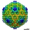
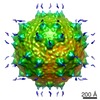
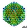
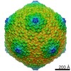
| Title | Three-dimensional icosahedral reconstruction of Rift Valley Fever virus at pH 7.4 | |||||||||
 Map data Map data | Rift Valley Fever virus at pH 7.4 Rift Valley fever Rift Valley fever | |||||||||
 Sample Sample |
| |||||||||
| Biological species |  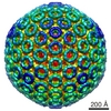 Rift Valley fever virus Rift Valley fever virus | |||||||||
| Method | single particle reconstruction / cryo EM / negative staining / Resolution: 20.0 Å | |||||||||
 Authors Authors | Huiskonen JT / Overby AK / Weber F / Gruenewald K | |||||||||
 Citation Citation | Journal: J Virol / Year: 2009 Title: Electron cryo-microscopy and single-particle averaging of Rift Valley fever virus: evidence for GN-GC glycoprotein heterodimers. Authors: Juha T Huiskonen / Anna K Overby / Friedemann Weber / Kay Grünewald /  Abstract: Rift Valley fever virus (RVFV) is a member of the genus Phlebovirus within the family Bunyaviridae. It is a mosquito-borne zoonotic agent that can cause hemorrhagic fever in humans. The enveloped ...Rift Valley fever virus (RVFV) is a member of the genus Phlebovirus within the family Bunyaviridae. It is a mosquito-borne zoonotic agent that can cause hemorrhagic fever in humans. The enveloped RVFV virions are known to be covered by capsomers of the glycoproteins G(N) and G(C), organized on a T=12 icosahedral lattice. However, the structural units forming the RVFV capsomers have not been determined. Conflicting biochemical results for another phlebovirus (Uukuniemi virus) have indicated the existence of either G(N) and G(C) homodimers or G(N)-G(C) heterodimers in virions. Here, we have studied the structure of RVFV using electron cryo-microscopy combined with three-dimensional reconstruction and single-particle averaging. The reconstruction at 2.2-nm resolution revealed the organization of the glycoprotein shell, the lipid bilayer, and a layer of ribonucleoprotein (RNP). Five- and six-coordinated capsomers are formed by the same basic structural unit. Molecular-mass measurements suggest a G(N)-G(C) heterodimer as the most likely candidate for this structural unit. Both leaflets of the lipid bilayer were discernible, and the glycoprotein transmembrane densities were seen to modulate the curvature of the lipid bilayer. RNP densities were situated directly underneath the transmembrane densities, suggesting an interaction between the glycoprotein cytoplasmic tails and the RNPs. The success of the single-particle averaging approach taken in this study suggests that it is applicable in the study of other phleboviruses, as well, enabling higher-resolution description of these medically important pathogens. | |||||||||
| History |
|
- Structure visualization
| Movie |
 Movie viewer Movie viewer |
|---|---|
| Structure viewer | EM map: SurfViewMolmilJmol/JSmol |
| Supplemental images |

- Downloads & links
-EMDB archive
| Map data | emd_1550.map.gz | 53.5 MB | EMDB map data format | |
|---|---|---|---|---|
| Header (meta data) | emd-1550-v30.xmlemd-1550.xml | 9.3 KB 9.3 KB | Display Display | EMDB header |
| Images | 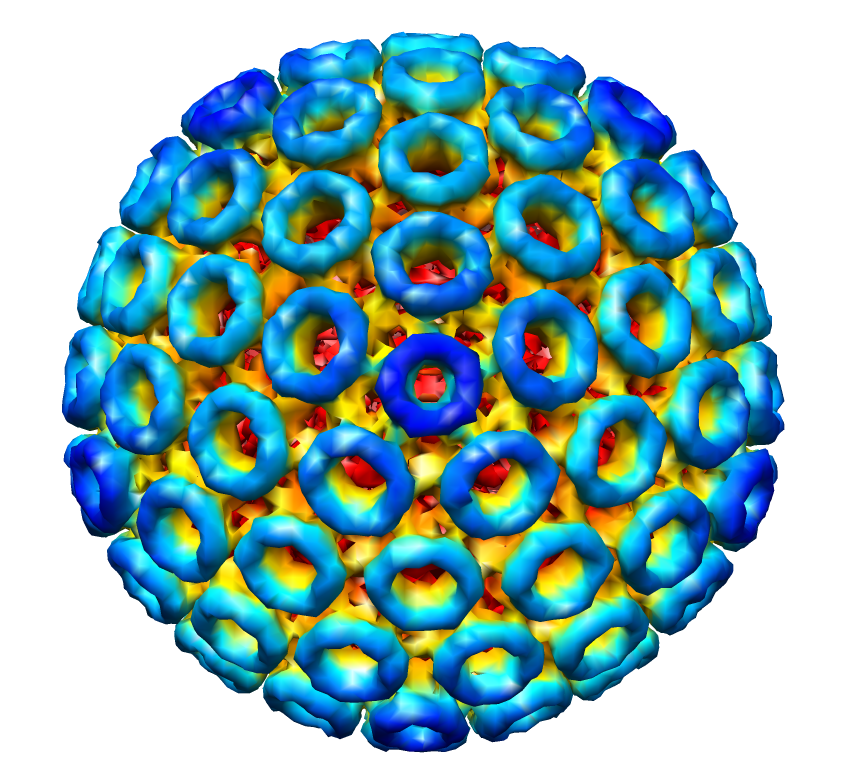 1550.png 1550.png | 601.9 KB | ||
| Archive directory |  http://ftp.pdbj.org/pub/emdb/structures/EMD-1550ftp://ftp.pdbj.org/pub/emdb/structures/EMD-1550 http://ftp.pdbj.org/pub/emdb/structures/EMD-1550ftp://ftp.pdbj.org/pub/emdb/structures/EMD-1550 | HTTPS FTP |
-Related structure data

-Links
| EMDB pages | EMDB (EBI/PDBe) / EMDataResource |
|---|
-Map
| File | Download / File: emd_1550.map.gz / Format: CCP4 / Size: 127.9 MB / Type: IMAGE STORED AS FLOATING POINT NUMBER (4 BYTES) | ||||||||||||||||||||||||||||||||||||||||||||||||||||||||||||||||||||
|---|---|---|---|---|---|---|---|---|---|---|---|---|---|---|---|---|---|---|---|---|---|---|---|---|---|---|---|---|---|---|---|---|---|---|---|---|---|---|---|---|---|---|---|---|---|---|---|---|---|---|---|---|---|---|---|---|---|---|---|---|---|---|---|---|---|---|---|---|---|
| Annotation | Rift Valley Fever virus at pH 7.4 | ||||||||||||||||||||||||||||||||||||||||||||||||||||||||||||||||||||
| Voxel size | X=Y=Z: 4 Å | ||||||||||||||||||||||||||||||||||||||||||||||||||||||||||||||||||||
| Density |
| ||||||||||||||||||||||||||||||||||||||||||||||||||||||||||||||||||||
| Symmetry | Space group: 1 | ||||||||||||||||||||||||||||||||||||||||||||||||||||||||||||||||||||
| Details | EMDB XML:
CCP4 map header:
| ||||||||||||||||||||||||||||||||||||||||||||||||||||||||||||||||||||
-Supplemental data
- Sample components
-Entire : Rift Valley Fever virus
| Entire | Name: Rift Valley Fever virus |
|---|---|
| Components |
|
-Supramolecule #1000: Rift Valley Fever virus
| Supramolecule | Name: Rift Valley Fever virus / type: sample / ID: 1000 Details: The sample was fixed with glutaraldehyde at pH 7.4 prior to virus purification Number unique components: 1 |
|---|
-Supramolecule #1: Rift Valley fever virus
| Supramolecule | Name: Rift Valley fever virus / type: virus / ID: 1 / Name.synonym: RVFV / Details: clone 13 / NCBI-ID: 11588 / Sci species name: Rift Valley fever virus / Virus type: VIRION / Virus isolate: STRAIN / Virus enveloped: Yes / Virus empty: No / Syn species name: RVFV |
|---|---|
| Host (natural) | Organism:  Ovis aries (sheep) / synonym: VERTEBRATES Ovis aries (sheep) / synonym: VERTEBRATES |
| Virus shell | Shell ID: 1 / Name: glycoprotein layer / Diameter: 1060 Å / T number (triangulation number): 12 |
-Experimental details
-Structure determination
| Method | negative staining, cryo EM |
|---|---|
 Processing Processing | single particle reconstruction |
| Aggregation state | particle |
-Sample preparation
| Buffer | pH: 7.4 / Details: 50 mM Tris-HCl pH 7.4, 100 mM NaCl |
|---|---|
| Staining | Type: NEGATIVE / Details: unstained sample |
| Grid | Details: 200 mesh copper C-flat |
| Vitrification | Cryogen name: ETHANE / Chamber temperature: 90 K / Instrument: HOMEMADE PLUNGER / Details: Vitrification instrument: MPI Biochemisty plunger / Method: Blot for 4 seconds before plunging |
- Electron microscopy
| Microscope | FEI TECNAI 20 |
|---|---|
| Electron beam | Acceleration voltage: 200 kV / Electron source: FIELD EMISSION GUN |
| Electron optics | Calibrated magnification: 112000 / Illumination mode: FLOOD BEAM / Imaging mode: BRIGHT FIELDBright-field microscopy / Cs: 2.0 mm / Nominal defocus max: 2.6 µm / Nominal defocus min: 1.2 µm / Nominal magnification: 80000 |
| Sample stage | Specimen holder: Side entry liquid nitrogen-cooled cryo specimen holder Specimen holder model: GATAN LIQUID NITROGEN |
| Temperature | Min: 90 K / Max: 90 K / Average: 90 K |
| Alignment procedure | Legacy - Astigmatism: objective lens astigmatism was corrected at 80,000 times magnification |
| Details | Low dose imaging |
| Date | Jun 30, 2008 |
| Image recording | Category: CCD / Film or detector model: FEI EAGLE (2k x 2k) / Digitization - Sampling interval: 15 µm / Number real images: 19 / Average electron dose: 20 e/Å2 / Bits/pixel: 16 |
| Tilt angle min | 0 |
| Tilt angle max | 0 |
-Image processing
| CTF correction | Details: Each image, phases flipped |
|---|---|
| Final reconstruction | Applied symmetry - Point group: I (icosahedral) / Algorithm: OTHER / Resolution.type: BY AUTHOR / Resolution: 20.0 Å / Resolution method: FSC 0.5 CUT-OFF / Software - Name: PFT2, EM3DR2 / Number images used: 113 |
| Details | The particles were selected using an automatic selection program ETHAN |
