 Movie
Movie Controller

[English] 日本語
 Yorodumi
- EMDB-1526: Single particle reconstruction of Methanothermobacter thermautotr... -
+ Open data

- Basic information
| Entry | Database: EMDB / ID: EMD-1526 | |||||||||
|---|---|---|---|---|---|---|---|---|---|---|
| Title | Single particle reconstruction of Methanothermobacter thermautotrophicus MCM protein bound to a 5,600bp dsDNA fragment. | |||||||||
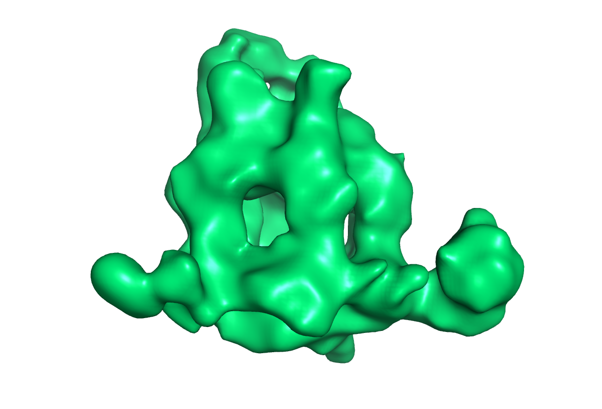
 Map data Map data | This is an electron density map of the MthMCM helicase treated with a 5,600 bp dsDNA fragment. | |||||||||
 Sample Sample |
| |||||||||
 Keywords Keywords | helicase / AAA+ ATPase / DNA replication | |||||||||
| Biological species | synthetic construct (others) /   Methanothermobacter thermautotrophicus str. Delta H (archaea) Methanothermobacter thermautotrophicus str. Delta H (archaea) | |||||||||
| Method | single particle reconstruction / cryo EM / Resolution: 19.0 Å | |||||||||
 Authors Authors | Costa A / van Duinen G / Medagli B / Chong J / Sakakibara N / Kelman Z / Nair SK / Patwardhan A / Onesti S | |||||||||
 Citation Citation | Journal: EMBO J / Year: 2008 Title: Cryo-electron microscopy reveals a novel DNA-binding site on the MCM helicase. Authors: Alessandro Costa / Gijs van Duinen / Barbara Medagli / James Chong / Nozomi Sakakibara / Zvi Kelman / Satish K Nair / Ardan Patwardhan / Silvia Onesti /  Abstract: The eukaryotic MCM2-7 complex is recruited at origins of replication during the G1 phase and acts as the main helicase at the replication fork during the S phase of the cell cycle. To characterize ...The eukaryotic MCM2-7 complex is recruited at origins of replication during the G1 phase and acts as the main helicase at the replication fork during the S phase of the cell cycle. To characterize the interplay between the MCM helicase and DNA prior to the melting of the double helix, we determined the structure of an archaeal MCM orthologue bound to a 5.6-kb double-stranded DNA segment, using cryo-electron microscopy. DNA wraps around the N-terminal face of a single hexameric ring. This interaction requires a conformational change within the outer belt of the MCM N-terminal domain, exposing a previously unrecognized helix-turn-helix DNA-binding motif. Our findings provide novel insights into the role of the MCM complex during the initiation step of DNA replication. | |||||||||
| History |
|
- Structure visualization
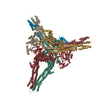
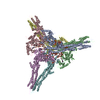
| Movie |
 Movie viewer Movie viewer |
|---|---|
| Structure viewer | EM map: SurfViewMolmilJmol/JSmol |
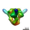
| Supplemental images |


- Downloads & links
-EMDB archive
| Map data | emd_1526.map.gz | 3.1 MB | EMDB map data format | |
|---|---|---|---|---|
| Header (meta data) | emd-1526-v30.xmlemd-1526.xml | 9.8 KB 9.8 KB | Display Display | EMDB header |
| Images |  1526.gif 1526.gif emd_1526.pngemd_1526.tif emd_1526.pngemd_1526.tif | 37.9 KB 507.6 KB 286.9 KB | ||
| Archive directory |  http://ftp.pdbj.org/pub/emdb/structures/EMD-1526ftp://ftp.pdbj.org/pub/emdb/structures/EMD-1526 http://ftp.pdbj.org/pub/emdb/structures/EMD-1526ftp://ftp.pdbj.org/pub/emdb/structures/EMD-1526 | HTTPS FTP |
-Validation report
| Summary document | emd_1526_validation.pdf.gz | 201.6 KB | Display | EMDB validaton report |
|---|---|---|---|---|
| Full document | emd_1526_full_validation.pdf.gz | 200.7 KB | Display | |
| Data in XML | emd_1526_validation.xml.gz | 5.3 KB | Display | |
| Arichive directory | https://ftp.pdbj.org/pub/emdb/validation_reports/EMD-1526ftp://ftp.pdbj.org/pub/emdb/validation_reports/EMD-1526 | HTTPS FTP |
-Related structure data
| Similar structure data |
|---|


-Links
| EMDB pages | EMDB (EBI/PDBe) / EMDataResource |
|---|
-Map
| File | Download / File: emd_1526.map.gz / Format: CCP4 / Size: 3.2 MB / Type: IMAGE STORED AS FLOATING POINT NUMBER (4 BYTES) | ||||||||||||||||||||||||||||||||||||||||||||||||||||||||||||||||||||
|---|---|---|---|---|---|---|---|---|---|---|---|---|---|---|---|---|---|---|---|---|---|---|---|---|---|---|---|---|---|---|---|---|---|---|---|---|---|---|---|---|---|---|---|---|---|---|---|---|---|---|---|---|---|---|---|---|---|---|---|---|---|---|---|---|---|---|---|---|---|
| Annotation | This is an electron density map of the MthMCM helicase treated with a 5,600 bp dsDNA fragment. | ||||||||||||||||||||||||||||||||||||||||||||||||||||||||||||||||||||
| Voxel size | X=Y=Z: 2.6 Å | ||||||||||||||||||||||||||||||||||||||||||||||||||||||||||||||||||||
| Density |
| ||||||||||||||||||||||||||||||||||||||||||||||||||||||||||||||||||||
| Symmetry | Space group: 1 | ||||||||||||||||||||||||||||||||||||||||||||||||||||||||||||||||||||
| Details | EMDB XML:
CCP4 map header:
| ||||||||||||||||||||||||||||||||||||||||||||||||||||||||||||||||||||
-Supplemental data
- Sample components
-Entire : Methanothermobacter thermautotrophicus MCM protein bound to a 5,6...
| Entire | Name: Methanothermobacter thermautotrophicus MCM protein bound to a 5,600 bp dsDNA fragment |
|---|---|
| Components |
|
-Supramolecule #1000: Methanothermobacter thermautotrophicus MCM protein bound to a 5,6...
| Supramolecule | Name: Methanothermobacter thermautotrophicus MCM protein bound to a 5,600 bp dsDNA fragment type: sample / ID: 1000 Oligomeric state: One homohexamer of MCM binds to one dsDNA fragment Number unique components: 2 |
|---|
-Macromolecule #1: DNA
| Macromolecule | Name: DNA / type: dna / ID: 1 / Name.synonym: 5,600 bp pET15b vector fragment / Classification: DNA / Structure: DOUBLE HELIX / Synthetic?: No |
|---|---|
| Source (natural) | Organism: synthetic construct (others) |
-Macromolecule #2: Mini-chromosome Maintenance complex
| Macromolecule | Name: Mini-chromosome Maintenance complex / type: protein_or_peptide / ID: 2 / Name.synonym: MCM / Number of copies: 6 / Oligomeric state: Hexamer / Recombinant expression: Yes |
|---|---|
| Source (natural) | Organism: Methanothermobacter thermautotrophicus str. Delta H (archaea) Strain: Delta H |
| Molecular weight | Theoretical: 450 KDa |
| Recombinant expression | Organism:  |
-Experimental details
-Structure determination
| Method | cryo EM |
|---|---|
 Processing Processing | single particle reconstruction |
| Aggregation state | particle |
-Sample preparation
| Concentration | 0.012 mg/mL |
|---|---|
| Buffer | pH: 7.5 Details: 30mM Tris HCl pH 7.5, 50 mM NaCl, 5mM MgCl2, 5 mM beta-mercaptoethanol |
| Grid | Details: Quantifoil R 1.2/1.3 |
| Vitrification | Cryogen name: ETHANE / Chamber humidity: 100 % / Chamber temperature: 4.5 K / Instrument: OTHER / Details: Vitrification instrument: FEI Vitrobot plunger Method: Blot for 2.5 seconds at an offset of -1 before plunging |
- Electron microscopy
| Microscope | FEI/PHILIPS CM200FEG |
|---|---|
| Image recording | Digitization - Scanner: OTHER / Digitization - Sampling interval: 6.5 µm / Number real images: 22 / Average electron dose: 17 e/Å2 |
| Electron beam | Acceleration voltage: 200 kV / Electron source:  FIELD EMISSION GUN FIELD EMISSION GUN |
| Electron optics | Illumination mode: SPOT SCAN / Imaging mode: BRIGHT FIELD / Nominal defocus max: 2.5 µm / Nominal defocus min: 0.7 µm / Nominal magnification: 50000 |
| Sample stage | Specimen holder: Side entry liquid nitrogen-cooled cryo specimen holder. Specimen holder model: GATAN LIQUID NITROGEN |
