 Movie
Movie Controller


+ Open data

- Basic information
| Entry | Database: EMDB / ID: EMD-1512 | |||||||||
|---|---|---|---|---|---|---|---|---|---|---|
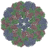
| Title | CryoEM structure of Blackcurrant Reversion Virus | |||||||||
 Map data Map data | This is a reconstruction of blackcurrant reversion virus at 17 angstrom resolution. | |||||||||
 Sample Sample |
| |||||||||
 Keywords Keywords | Cryo / electron / microscopy / structure / blackcurrant / reversion / virus | |||||||||
| Biological species |  Blackcurrant reversion virus Blackcurrant reversion virus | |||||||||
| Method | single particle reconstruction / cryo EM / negative staining / Resolution: 17.0 Å | |||||||||
 Authors Authors | Seitsonen JJT / Susi P / Lemmetty A / Butcher SJ | |||||||||
 Citation Citation | Journal: Virology / Year: 2008 Title: Structure of the mite-transmitted Blackcurrant reversion nepovirus using electron cryo-microscopy. Authors: Jani J T Seitsonen / Petri Susi / Anne Lemmetty / Sarah J Butcher /  Abstract: Blackcurrant reversion nepovirus (BRV; genus Nepovirus) has a single-stranded, bipartite RNA genome surrounded by 60 copies of a single capsid protein (CP). BRV is the most important mite-transmitted ...Blackcurrant reversion nepovirus (BRV; genus Nepovirus) has a single-stranded, bipartite RNA genome surrounded by 60 copies of a single capsid protein (CP). BRV is the most important mite-transmitted viral pathogen of the Ribes species. It is the causal agent of blackcurrant reversion disease. We determined the structure of BRV to 1.7 nm resolution using electron cryo- microscopy (cryoEM) and image reconstruction. The reconstruction reveals a pseudo T=3 viral capsid similar to that of tobacco ringspot virus (TRSV). We modelled the BRV capsid protein to that of TRSV and fitted it into the cryoEM reconstruction. The fit indicated that the extended C-terminus of BRV-CP is located on the capsid surface and the N-terminus on the interior. We generated peptide antibodies to two putatively exposed C-terminal sequences and these reacted with the virus. Hence homology modelling may be useful for defining epitopes for antibody generation for diagnostic testing of BRV in commercial crops. | |||||||||
| History |
|
- Structure visualization
| Movie |
 Movie viewer Movie viewer |
|---|---|
| Structure viewer | EM map: SurfViewMolmilJmol/JSmol |
| Supplemental images |

- Downloads & links
-EMDB archive
| Map data | emd_1512.map.gz | 4 MB | EMDB map data format | |
|---|---|---|---|---|
| Header (meta data) | emd-1512-v30.xmlemd-1512.xml | 9.9 KB 9.9 KB | Display Display | EMDB header |
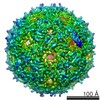
| Images | 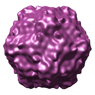 1512.gif 1512.gif | 75.4 KB | ||
| Archive directory |  http://ftp.pdbj.org/pub/emdb/structures/EMD-1512ftp://ftp.pdbj.org/pub/emdb/structures/EMD-1512 http://ftp.pdbj.org/pub/emdb/structures/EMD-1512ftp://ftp.pdbj.org/pub/emdb/structures/EMD-1512 | HTTPS FTP |
-Validation report
| Summary document | emd_1512_validation.pdf.gz | 203.3 KB | Display | EMDB validaton report |
|---|---|---|---|---|
| Full document | emd_1512_full_validation.pdf.gz | 202.5 KB | Display | |
| Data in XML | emd_1512_validation.xml.gz | 6.7 KB | Display | |
| Arichive directory | https://ftp.pdbj.org/pub/emdb/validation_reports/EMD-1512ftp://ftp.pdbj.org/pub/emdb/validation_reports/EMD-1512 | HTTPS FTP |
-Related structure data
| Similar structure data |
|---|

-Links
| EMDB pages | EMDB (EBI/PDBe) / EMDataResource |
|---|
-Map
| File | Download / File: emd_1512.map.gz / Format: CCP4 / Size: 30.3 MB / Type: IMAGE STORED AS FLOATING POINT NUMBER (4 BYTES) | ||||||||||||||||||||||||||||||||||||||||||||||||||||||||||||||||||||
|---|---|---|---|---|---|---|---|---|---|---|---|---|---|---|---|---|---|---|---|---|---|---|---|---|---|---|---|---|---|---|---|---|---|---|---|---|---|---|---|---|---|---|---|---|---|---|---|---|---|---|---|---|---|---|---|---|---|---|---|---|---|---|---|---|---|---|---|---|---|
| Annotation | This is a reconstruction of blackcurrant reversion virus at 17 angstrom resolution. | ||||||||||||||||||||||||||||||||||||||||||||||||||||||||||||||||||||
| Voxel size | X=Y=Z: 2.26203 Å | ||||||||||||||||||||||||||||||||||||||||||||||||||||||||||||||||||||
| Density |
| ||||||||||||||||||||||||||||||||||||||||||||||||||||||||||||||||||||
| Symmetry | Space group: 1 | ||||||||||||||||||||||||||||||||||||||||||||||||||||||||||||||||||||
| Details | EMDB XML:
CCP4 map header:
| ||||||||||||||||||||||||||||||||||||||||||||||||||||||||||||||||||||
-Supplemental data
- Sample components
-Entire : Purified blackcurrant reversion virus in 0.05 M Na-citrate buffer...
| Entire | Name: Purified blackcurrant reversion virus in 0.05 M Na-citrate buffer (pH 7.0) |
|---|---|
| Components |
|
-Supramolecule #1000: Purified blackcurrant reversion virus in 0.05 M Na-citrate buffer...
| Supramolecule | Name: Purified blackcurrant reversion virus in 0.05 M Na-citrate buffer (pH 7.0) type: sample / ID: 1000 Details: The sample was stored at -80 degrees celcius and thawed before loading onto grids Oligomeric state: Complete particle, (60-mer) with the RNA genome inside Number unique components: 1 |
|---|---|
| Molecular weight | Experimental: 3.30 MDa / Theoretical: 3.45 MDa Method: SDS-PAGE (55 kDa per capsid protein, 60 capsid proteins per capsid) (Lemmetty et al. 1997, The American Phytopathology Society) |
-Supramolecule #1: Blackcurrant reversion virus
| Supramolecule | Name: Blackcurrant reversion virus / type: virus / ID: 1 / Name.synonym: BRV / NCBI-ID: 65743 / Sci species name: Blackcurrant reversion virus / Virus type: VIRION / Virus isolate: SPECIES / Virus enveloped: No / Virus empty: No / Syn species name: BRV |
|---|---|
| Host (natural) | Organism:  Chenopodium quinoa (quinoa) / synonym: PLANTAE(HIGHER PLANTS) Chenopodium quinoa (quinoa) / synonym: PLANTAE(HIGHER PLANTS) |
| Molecular weight | Experimental: 3.30 MDa / Theoretical: 3.45 MDa |
| Virus shell | Shell ID: 1 / Name: Coat protein / Diameter: 290 Å / T number (triangulation number): 1 |
-Experimental details
-Structure determination
| Method | negative staining, cryo EM |
|---|---|
 Processing Processing | single particle reconstruction |
| Aggregation state | particle |
-Sample preparation
| Concentration | 1 mg/mL |
|---|---|
| Buffer | pH: 7 / Details: 0.05 M Na-citrate buffer |
| Staining | Type: NEGATIVE Details: Vitrified. Grids were blotted for roughly one second before being plunged into liquid ethane. |
| Grid | Details: C-Flat 224 grids |
| Vitrification | Cryogen name: ETHANE / Instrument: HOMEMADE PLUNGER / Details: Vitrification instrument: Guillotine / Method: Blot for 1 second before plunging |
- Electron microscopy
| Microscope | FEI TECNAI F20 |
|---|---|
| Alignment procedure | Legacy - Astigmatism: astigmatism was corrected at 62000x magnification |
| Details | Part of the data was recorded on Kodak SO163 film at nominal magnification of 50000. |
| Image recording | Category: CCD / Film or detector model: GATAN ULTRASCAN 4000 (4k x 4k) / Digitization - Sampling interval: 15 µm / Number real images: 65 / Average electron dose: 17 e/Å2 Details: 65 images total, 10 images were scanned from film using Zeiss Photoscan TD scanner with a stepsize of 7 micrometers. |
| Electron beam | Acceleration voltage: 200 kV / Electron source:  FIELD EMISSION GUN FIELD EMISSION GUN |
| Electron optics | Calibrated magnification: 66400 / Illumination mode: SPOT SCAN / Imaging mode: BRIGHT FIELD / Cs: 2.0 mm / Nominal defocus max: 4.6 µm / Nominal defocus min: 1.6 µm / Nominal magnification: 68000 |
| Sample stage | Specimen holder: Gatan 626 / Specimen holder model: GATAN LIQUID NITROGEN |
| Experimental equipment |  Model: Tecnai F20 / Image courtesy: FEI Company |
-Image processing
| CTF correction | Details: each micrograph |
|---|---|
| Final reconstruction | Applied symmetry - Point group: I (icosahedral) / Algorithm: OTHER / Resolution.type: BY AUTHOR / Resolution: 17.0 Å / Resolution method: FSC 0.5 CUT-OFF / Software - Name: PFT, POR, EM3DR, P3DR Details: Particles from CCD data and film data were combined for the final reconstruction. Number images used: 831 |
