 Movie
Movie Controller

[English] 日本語
 Yorodumi
- EMDB-1463: Three-dimensional structure of canine adenovirus serotype 2 capsid -
+ Open data

- Basic information
| Entry | Database: EMDB / ID: EMD-1463 | |||||||||
|---|---|---|---|---|---|---|---|---|---|---|
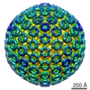
| Title | Three-dimensional structure of canine adenovirus serotype 2 capsid | |||||||||
 Map data Map data | Density map of canine adenovirus with C-terminal of protein IX fused with GFP protein | |||||||||
 Sample Sample |
| |||||||||
| Biological species |   Canine adenovirus 2 Canine adenovirus 2 | |||||||||
| Method | single particle reconstruction / cryo EM / negative staining / Resolution: 25.0 Å | |||||||||
 Authors Authors | Schoehn G / El Bakkouri M / Fabry CM / Billet O / Estrozi LF / Le L / Curiel DT / Kajava AV / Ruigrok RW / Kremer EJ | |||||||||
 Citation Citation | Journal: J Virol / Year: 2008 Title: Three-dimensional structure of canine adenovirus serotype 2 capsid. Authors: Guy Schoehn / Majida El Bakkouri / Céline M S Fabry / Oliver Billet / Leandro F Estrozi / Long Le / David T Curiel / Andrey V Kajava / Rob W H Ruigrok / Eric J Kremer /  Abstract: There are more than 100 known adenovirus (AdV) serotypes, including 50 human serotypes. Because AdV-induced disease is relatively species specific, vectors derived from nonhuman serotypes may have ...There are more than 100 known adenovirus (AdV) serotypes, including 50 human serotypes. Because AdV-induced disease is relatively species specific, vectors derived from nonhuman serotypes may have wider clinical potential based, in part, on the lack of ubiquitous memory immunity. Whereas a few of the human serotype capsids have been studied at the structural level, none of the nonhuman serotypes has been analyzed. The basis laid by the analysis of human AdV (hAdV) has allowed us to determine and compare the three-dimensional structure of the capsid of canine serotype 2 (CAV-2) to that of hAdV serotype 5 (hAdV-5). We show that CAV-2 capsid has a smoother structure than the human serotypes. Many of the external loops found in the hAdV-5 penton base and the hexon, against which the antibody response is directed, are shorter or absent in CAV-2. On the other hand, the CAV-2 fiber appears to be more complex, with two bends in the shaft. An interesting difference between the human and canine viruses is that the C-terminal part of protein IX is in a different position, making an antenna sticking out of the CAV-2 capsid. The comparison between the two viruses allows the identification of sites that should be easy to modify on the CAV-2 capsid for altering tissue tropism or other biological activities. | |||||||||
| History |
|
- Structure visualization
| Movie |
 Movie viewer Movie viewer |
|---|---|
| Structure viewer | EM map: SurfViewMolmilJmol/JSmol |
| Supplemental images |

- Downloads & links
-EMDB archive
| Map data | emd_1463.map.gz | 99.1 MB | EMDB map data format | |
|---|---|---|---|---|
| Header (meta data) | emd-1463-v30.xmlemd-1463.xml | 9 KB 9 KB | Display Display | EMDB header |
| Images | 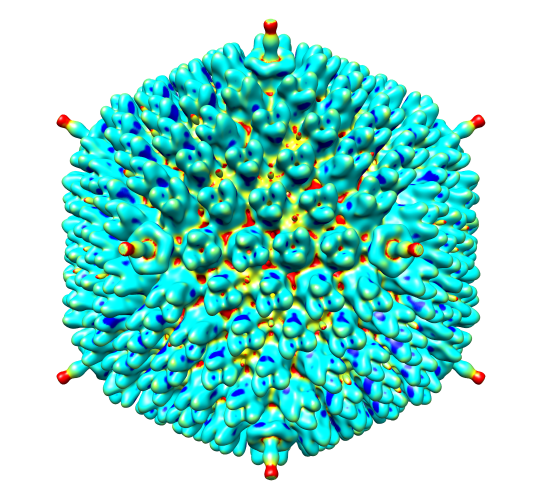 emd_1463.png emd_1463.png | 321.9 KB | ||
| Archive directory |  http://ftp.pdbj.org/pub/emdb/structures/EMD-1463ftp://ftp.pdbj.org/pub/emdb/structures/EMD-1463 http://ftp.pdbj.org/pub/emdb/structures/EMD-1463ftp://ftp.pdbj.org/pub/emdb/structures/EMD-1463 | HTTPS FTP |
-Related structure data


-Links
| EMDB pages | EMDB (EBI/PDBe) / EMDataResource |
|---|
-Map
| File | Download / File: emd_1463.map.gz / Format: CCP4 / Size: 332.7 MB / Type: IMAGE STORED AS FLOATING POINT NUMBER (4 BYTES) | ||||||||||||||||||||||||||||||||||||||||||||||||||||||||||||||||||||
|---|---|---|---|---|---|---|---|---|---|---|---|---|---|---|---|---|---|---|---|---|---|---|---|---|---|---|---|---|---|---|---|---|---|---|---|---|---|---|---|---|---|---|---|---|---|---|---|---|---|---|---|---|---|---|---|---|---|---|---|---|---|---|---|---|---|---|---|---|---|
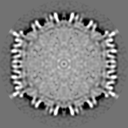
| Annotation | Density map of canine adenovirus with C-terminal of protein IX fused with GFP protein | ||||||||||||||||||||||||||||||||||||||||||||||||||||||||||||||||||||
| Projections & slices | Image control
Images are generated by Spider. | ||||||||||||||||||||||||||||||||||||||||||||||||||||||||||||||||||||
| Voxel size | X=Y=Z: 2.54 Å | ||||||||||||||||||||||||||||||||||||||||||||||||||||||||||||||||||||
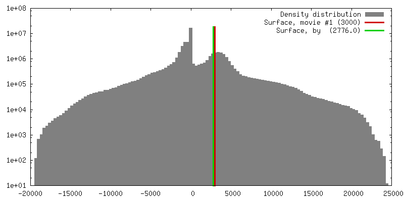
| Density |
| ||||||||||||||||||||||||||||||||||||||||||||||||||||||||||||||||||||
| Symmetry | Space group: 1 | ||||||||||||||||||||||||||||||||||||||||||||||||||||||||||||||||||||
| Details | EMDB XML:
CCP4 map header:
| ||||||||||||||||||||||||||||||||||||||||||||||||||||||||||||||||||||
 Z (Sec.)
 Y (Row.)
 X (Col.)


-Supplemental data
- Sample components
-Entire : Canine adenovirus serotype 2
| Entire | Name: Canine adenovirus serotype 2 |
|---|---|
| Components |
|
-Supramolecule #1000: Canine adenovirus serotype 2
| Supramolecule | Name: Canine adenovirus serotype 2 / type: sample / ID: 1000 Details: The protein IX is fused with the GFP protein at his C termini Number unique components: 1 |
|---|---|
| Molecular weight | Experimental: 150 MDa |
-Supramolecule #1: Canine adenovirus 2
| Supramolecule | Name: Canine adenovirus 2 / type: virus / ID: 1 / NCBI-ID: 10514 / Sci species name: Canine adenovirus 2 / Virus type: VIRION / Virus isolate: SEROTYPE / Virus enveloped: No / Virus empty: No |
|---|---|
| Host (natural) | Organism:  Canis lupus familiaris (dog) / synonym: VERTEBRATES Canis lupus familiaris (dog) / synonym: VERTEBRATES |
| Virus shell | Shell ID: 1 / Name: Capsid / Diameter: 100 Å / T number (triangulation number): 25 |
-Experimental details
-Structure determination
| Method | negative staining, cryo EM |
|---|---|
 Processing Processing | single particle reconstruction |
| Aggregation state | particle |
-Sample preparation
| Concentration | 1 mg/mL |
|---|---|
| Buffer | pH: 7.4 / Details: Tris ph 7.4 NaCl 150 mM |
| Staining | Type: NEGATIVE / Details: Cryo EM |
| Grid | Details: Quantifoil |
| Vitrification | Cryogen name: ETHANE / Instrument: OTHER / Details: Vitrification instrument: Zeiss / Method: Blot for 2 seconds before plunging |
- Electron microscopy
| Microscope | FEI/PHILIPS CM200T |
|---|---|
| Electron beam | Acceleration voltage: 200 kV / Electron source: LAB6 |
| Electron optics | Illumination mode: OTHER / Imaging mode: BRIGHT FIELDBright-field microscopy / Cs: 2 mm / Nominal defocus max: 3.0 µm / Nominal defocus min: 1.0 µm / Nominal magnification: 27500 |
| Sample stage | Specimen holder: Eucentric / Specimen holder model: GATAN LIQUID NITROGEN |
| Alignment procedure | Legacy - Astigmatism: Objective lens astigmatism was corrected at 100,000 times |
| Image recording | Category: FILM / Film or detector model: KODAK SO-163 FILM / Digitization - Scanner: ZEISS SCAI / Digitization - Sampling interval: 7 µm / Number real images: 20 / Bits/pixel: 8 |
-Image processing
| CTF correction | Details: Each negative |
|---|---|
| Final reconstruction | Applied symmetry - Point group: I (icosahedral) / Algorithm: OTHER / Resolution.type: BY AUTHOR / Resolution: 25.0 Å / Resolution method: FSC 0.5 CUT-OFF / Software - Name: PFT2, EM3DR2 / Number images used: 800 |
