 Movie
Movie Controller


+ Open data

- Basic information
| Entry | Database: EMDB / ID: EMD-1415 | |||||||||
|---|---|---|---|---|---|---|---|---|---|---|
| Title | Cryo-EM study of the Pseudomonas bacteriophage phiKZ. | |||||||||
 Map data Map data | This is a 3D model of the bacteriophage phiKZ based on the previously deposited reconstructions of the capsid (EMD-1392) and the tail (EMD-1393) | |||||||||
 Sample Sample |
| |||||||||
| Biological species |  Pseudomonas phage phiKZ (virus) Pseudomonas phage phiKZ (virus) | |||||||||
| Method |  single particle reconstruction / cryo EM / Resolution: 28.0 Å single particle reconstruction / cryo EM / Resolution: 28.0 Å | |||||||||
 Authors Authors | Fokine A / Battisti A / Bowman V / Efimov A / Kurochkina L / Chipman P / Mesyanzhinov V / Rossmann M | |||||||||
 Citation Citation | Journal: Structure / Year: 2007 Title: Cryo-EM study of the Pseudomonas bacteriophage phiKZ. Authors: Andrei Fokine / Anthony J Battisti / Valorie D Bowman / Andrei V Efimov / Lidia P Kurochkina / Paul R Chipman / Vadim V Mesyanzhinov / Michael G Rossmann /  Abstract: The phiKZ virus is one of the largest known bacteriophages. It infects Pseudomonas aeruginosa, which is frequently pathogenic in humans, and, therefore, has potential for phage therapy. The phiKZ ...The phiKZ virus is one of the largest known bacteriophages. It infects Pseudomonas aeruginosa, which is frequently pathogenic in humans, and, therefore, has potential for phage therapy. The phiKZ virion consists of an approximately 1450 A diameter icosahedral head and an approximately 2000 A long contractile tail. The structure of the phiKZ tail has been determined using cryo-electron microscopy. The phiKZ tail is much longer than that of bacteriophage T4. However, the helical parameters of their contractile sheaths, surrounding their tail tubes, are comparable. Although there is no recognizable sequence similarity between the phiKZ and T4 tail sheath proteins, they are similar in size and shape, suggesting that they evolved from a common ancestor. The phiKZ baseplate is significantly larger than that of T4 and has a flatter shape. Nevertheless, phiKZ, similar to T4, has a cell-puncturing device in the middle of its baseplate. | |||||||||
| History |
|
- Structure visualization
| Movie |
 Movie viewer Movie viewer |
|---|---|
| Structure viewer | EM map: SurfViewMolmilJmol/JSmol |
| Supplemental images |

- Downloads & links
-EMDB archive
| Map data | emd_1415.map.gz | 12.3 MB | EMDB map data format | |
|---|---|---|---|---|
| Header (meta data) | emd-1415-v30.xmlemd-1415.xml | 8.8 KB 8.8 KB | Display Display | EMDB header |
| Images | 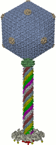 1415.gif 1415.gif | 35.5 KB | ||
| Archive directory |  http://ftp.pdbj.org/pub/emdb/structures/EMD-1415ftp://ftp.pdbj.org/pub/emdb/structures/EMD-1415 http://ftp.pdbj.org/pub/emdb/structures/EMD-1415ftp://ftp.pdbj.org/pub/emdb/structures/EMD-1415 | HTTPS FTP |
-Related structure data

-Links
| EMDB pages | EMDB (EBI/PDBe) / EMDataResource |
|---|
-Map
| File | Download / File: emd_1415.map.gz / Format: CCP4 / Size: 48.3 MB / Type: IMAGE STORED AS FLOATING POINT NUMBER (4 BYTES) | ||||||||||||||||||||||||||||||||||||||||||||||||||||||||||||||||||||
|---|---|---|---|---|---|---|---|---|---|---|---|---|---|---|---|---|---|---|---|---|---|---|---|---|---|---|---|---|---|---|---|---|---|---|---|---|---|---|---|---|---|---|---|---|---|---|---|---|---|---|---|---|---|---|---|---|---|---|---|---|---|---|---|---|---|---|---|---|---|
| Annotation | This is a 3D model of the bacteriophage phiKZ based on the previously deposited reconstructions of the capsid (EMD-1392) and the tail (EMD-1393) | ||||||||||||||||||||||||||||||||||||||||||||||||||||||||||||||||||||
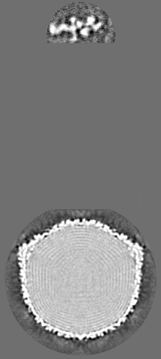
| Projections & slices | Image control
Images are generated by Spider. generated in cubic-lattice coordinate | ||||||||||||||||||||||||||||||||||||||||||||||||||||||||||||||||||||
| Voxel size | X=Y=Z: 8.4848 Å | ||||||||||||||||||||||||||||||||||||||||||||||||||||||||||||||||||||
| Density |
| ||||||||||||||||||||||||||||||||||||||||||||||||||||||||||||||||||||
| Symmetry | Space group: 1 | ||||||||||||||||||||||||||||||||||||||||||||||||||||||||||||||||||||
| Details | EMDB XML:
CCP4 map header:
| ||||||||||||||||||||||||||||||||||||||||||||||||||||||||||||||||||||
 Z (Sec.)
 Y (Row.)
 X (Col.)


-Supplemental data
- Sample components
-Entire : Bacteriophage phiKZ
| Entire | Name: Bacteriophage phiKZ |
|---|---|
| Components |
|
-Supramolecule #1000: Bacteriophage phiKZ
| Supramolecule | Name: Bacteriophage phiKZ / type: sample / ID: 1000 / Oligomeric state: one / Number unique components: 1 |
|---|
-Supramolecule #1: Pseudomonas phage phiKZ
| Supramolecule | Name: Pseudomonas phage phiKZ / type: virus / ID: 1 / NCBI-ID: 169683 / Sci species name: Pseudomonas phage phiKZ / Database: NCBI / Virus type: VIRION / Virus isolate: STRAIN / Virus enveloped: No / Virus empty: No |
|---|---|
| Host (natural) | Organism:   Pseudomonas aeruginosa (bacteria) / synonym: BACTERIA(EUBACTERIA) Pseudomonas aeruginosa (bacteria) / synonym: BACTERIA(EUBACTERIA) |
-Experimental details
-Structure determination
| Method | cryo EM |
|---|---|
 Processing Processing | single particle reconstruction |
| Aggregation state | particle |
-Sample preparation
| Buffer | pH: 7.5 / Details: 50 mM tris-Acetate-EDTA buffer (pH 7.5) |
|---|---|
| Vitrification | Cryogen name: ETHANE / Chamber humidity: 40 % / Instrument: HOMEMADE PLUNGER / Details: Vitrification instrument: in house manufactured / Method: hand blot 3 seconds, plugging during blot |
- Electron microscopy
| Microscope | FEI/PHILIPS CM300FEG/T |
|---|---|
| Electron beam | Acceleration voltage: 300 kV / Electron source: FIELD EMISSION GUN |
| Electron optics | Calibrated magnification: 33200 / Illumination mode: FLOOD BEAM / Imaging mode: BRIGHT FIELDBright-field microscopy / Cs: 2.0 mm / Nominal defocus max: 3.2 µm / Nominal defocus min: 0.9 µm / Nominal magnification: 33000 |
| Sample stage | Specimen holder model: GATAN LIQUID NITROGEN |
| Alignment procedure | Legacy - Astigmatism: live fft |
| Image recording | Category: FILM / Film or detector model: KODAK SO-163 FILM / Digitization - Scanner: ZEISS SCAI / Digitization - Sampling interval: 7 µm / Number real images: 112 / Average electron dose: 20 e/Å2 / Bits/pixel: 8 |
-Image processing
| Final reconstruction | Resolution.type: BY AUTHOR / Resolution: 28.0 Å / Resolution method: OTHER Details: This is a model of the phiKZ virion based on the previously deposited reconstructions of the phiKZ capsid and the tail part. |
|---|
