 Movie
Movie Controller

[English] 日本語
 Yorodumi
- EMDB-1411: Interaction of decay-accelerating factor with coxsackievirus B3. -
+ Open data

- Basic information
| Entry | Database: EMDB / ID: EMD-1411 | |||||||||
|---|---|---|---|---|---|---|---|---|---|---|
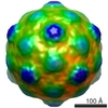
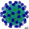
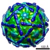
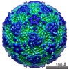
| Title | Interaction of decay-accelerating factor with coxsackievirus B3. | |||||||||
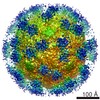
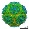
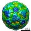
 Map data Map data | surface rendered coxsackievirus B3, M strain | |||||||||
 Sample Sample |
| |||||||||
| Biological species |  Coxsackievirus Coxsackievirus | |||||||||
| Method | single particle reconstruction / cryo EM / negative staining / Resolution: 27.0 Å | |||||||||
 Authors Authors | Hafenstein S | |||||||||
 Citation Citation | Journal: J Virol / Year: 2007 Title: Interaction of decay-accelerating factor with coxsackievirus B3. Authors: Susan Hafenstein / Valorie D Bowman / Paul R Chipman / Carol M Bator Kelly / Feng Lin / M Edward Medof / Michael G Rossmann /  Abstract: Many entero-, parecho-, and rhinoviruses use immunoglobulin (Ig)-like receptors that bind into the viral canyon and are required to initiate viral uncoating during infection. However, some of these ...Many entero-, parecho-, and rhinoviruses use immunoglobulin (Ig)-like receptors that bind into the viral canyon and are required to initiate viral uncoating during infection. However, some of these viruses use an alternative or additional receptor that binds outside the canyon. Both the coxsackievirus-adenovirus receptor (CAR), an Ig-like molecule that binds into the viral canyon, and decay-accelerating factor (DAF) have been identified as cellular receptors for coxsackievirus B3 (CVB3). A cryoelectron microscopy reconstruction of a variant of CVB3 complexed with DAF shows full occupancy of the DAF receptor in each of 60 binding sites. The DAF molecule bridges the canyon, blocking the CAR binding site and causing the two receptors to compete with one another. The binding site of DAF on CVB3 differs from the binding site of DAF on the surface of echoviruses, suggesting independent evolutionary processes. | |||||||||
| History |
|
- Structure visualization
| Movie |
 Movie viewer Movie viewer |
|---|---|
| Structure viewer | EM map: SurfViewMolmilJmol/JSmol |
| Supplemental images |
 UCSF Chimera
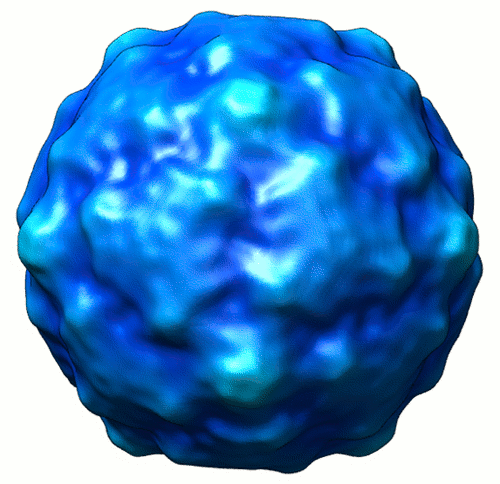
- Downloads & links
-EMDB archive
| Map data | emd_1411.map.gz | 8.5 MB | EMDB map data format | |
|---|---|---|---|---|
| Header (meta data) | emd-1411-v30.xmlemd-1411.xml | 9.7 KB 9.7 KB | Display Display | EMDB header |
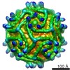
| Images |  1411.gif 1411.gif | 116.5 KB | ||
| Archive directory |  http://ftp.pdbj.org/pub/emdb/structures/EMD-1411ftp://ftp.pdbj.org/pub/emdb/structures/EMD-1411 http://ftp.pdbj.org/pub/emdb/structures/EMD-1411ftp://ftp.pdbj.org/pub/emdb/structures/EMD-1411 | HTTPS FTP |
-Validation report
| Summary document | emd_1411_validation.pdf.gz | 217.7 KB | Display | EMDB validaton report |
|---|---|---|---|---|
| Full document | emd_1411_full_validation.pdf.gz | 216.9 KB | Display | |
| Data in XML | emd_1411_validation.xml.gz | 6.4 KB | Display | |
| Arichive directory | https://ftp.pdbj.org/pub/emdb/validation_reports/EMD-1411ftp://ftp.pdbj.org/pub/emdb/validation_reports/EMD-1411 | HTTPS FTP |
-Related structure data

-Links
| EMDB pages | EMDB (EBI/PDBe) / EMDataResource |
|---|
-Map
| File | Download / File: emd_1411.map.gz / Format: CCP4 / Size: 22.1 MB / Type: IMAGE STORED AS SIGNED INTEGER (2 BYTES) | ||||||||||||||||||||||||||||||||||||||||||||||||||||||||||||||||||||
|---|---|---|---|---|---|---|---|---|---|---|---|---|---|---|---|---|---|---|---|---|---|---|---|---|---|---|---|---|---|---|---|---|---|---|---|---|---|---|---|---|---|---|---|---|---|---|---|---|---|---|---|---|---|---|---|---|---|---|---|---|---|---|---|---|---|---|---|---|---|
| Annotation | surface rendered coxsackievirus B3, M strain | ||||||||||||||||||||||||||||||||||||||||||||||||||||||||||||||||||||
| Voxel size | X=Y=Z: 2.22 Å | ||||||||||||||||||||||||||||||||||||||||||||||||||||||||||||||||||||
| Density |
| ||||||||||||||||||||||||||||||||||||||||||||||||||||||||||||||||||||
| Symmetry | Space group: 1 | ||||||||||||||||||||||||||||||||||||||||||||||||||||||||||||||||||||
| Details | EMDB XML:
CCP4 map header:
| ||||||||||||||||||||||||||||||||||||||||||||||||||||||||||||||||||||
-Supplemental data
- Sample components
-Entire : coxsackievirus B3, M strain
| Entire | Name: coxsackievirus B3, M strain |
|---|---|
| Components |
|
-Supramolecule #1000: coxsackievirus B3, M strain
| Supramolecule | Name: coxsackievirus B3, M strain / type: sample / ID: 1000 / Number unique components: 1 |
|---|
-Supramolecule #1: Coxsackievirus
| Supramolecule | Name: Coxsackievirus / type: virus / ID: 1 / Name.synonym: cvb3 / Details: Native virus control / NCBI-ID: 12066 / Sci species name: Coxsackievirus / Virus type: VIRION / Virus isolate: STRAIN / Virus enveloped: No / Virus empty: No / Syn species name: cvb3 |
|---|---|
| Host (natural) | Organism:  Homo sapiens (human) / synonym: VERTEBRATES Homo sapiens (human) / synonym: VERTEBRATES |
| Molecular weight | Experimental: 7.0 MDa / Theoretical: 7.0 MDa |
| Virus shell | Shell ID: 1 / Name: VP1 / Diameter: 300 Å / T number (triangulation number): 1 |
| Virus shell | Shell ID: 2 / Name: VP2 / Diameter: 300 Å / T number (triangulation number): 1 |
| Virus shell | Shell ID: 3 / Name: VP3 / Diameter: 300 Å / T number (triangulation number): 1 |
-Experimental details
-Structure determination
| Method | negative staining, cryo EM |
|---|---|
 Processing Processing | single particle reconstruction |
| Aggregation state | particle |
-Sample preparation
| Concentration | 2 mg/mL |
|---|---|
| Buffer | pH: 6 / Details: 50mM MES, pH 6.0 |
| Staining | Type: NEGATIVE Details: small aliquotes were applied to grids and vitrified |
| Vitrification | Cryogen name: ETHANE / Chamber temperature: 120 K / Instrument: HOMEMADE PLUNGER / Details: Vitrification instrument: plunger / Method: blot before plunging |
- Electron microscopy
| Microscope | FEI/PHILIPS CM300FEG/T |
|---|---|
| Temperature | Min: 83 K / Max: 83 K / Average: 93 K |
| Alignment procedure | Legacy - Astigmatism: lens astigmatism was corrected at 98,000 times mag |
| Date | Aug 23, 2000 |
| Image recording | Category: FILM / Film or detector model: KODAK SO-163 FILM / Digitization - Scanner: ZEISS SCAI / Digitization - Sampling interval: 7 µm / Number real images: 9 / Average electron dose: 37 e/Å2 / Details: scanned at 7 microns and bin averaged to 14 / Od range: 0.9 / Bits/pixel: 8 |
| Tilt angle min | 0 |
| Tilt angle max | 0 |
| Electron beam | Acceleration voltage: 300 kV / Electron source: TUNGSTEN HAIRPIN |
| Electron optics | Calibrated magnification: 63100 / Illumination mode: FLOOD BEAM / Imaging mode: BRIGHT FIELD / Cs: 2.0 mm / Nominal defocus max: 3.5 µm / Nominal defocus min: 0.7 µm / Nominal magnification: 61000 |
| Sample stage | Specimen holder: side mounted nitrogen cooled / Specimen holder model: GATAN LIQUID NITROGEN |
-Image processing
| CTF correction | Details: robem |
|---|---|
| Final reconstruction | Applied symmetry - Point group: I (icosahedral) / Algorithm: OTHER / Resolution.type: BY AUTHOR / Resolution: 27.0 Å / Resolution method: FSC 0.5 CUT-OFF / Software - Name: EMPFT, EM3DR / Number images used: 600 |
