 Movie
Movie Controller

[English] 日本語
 Yorodumi
- EMDB-1339: Genome sequence, structural proteins, and capsid organization of ... -
+ Open data

- Basic information
| Entry | Database: EMDB / ID: EMD-1339 | |||||||||
|---|---|---|---|---|---|---|---|---|---|---|
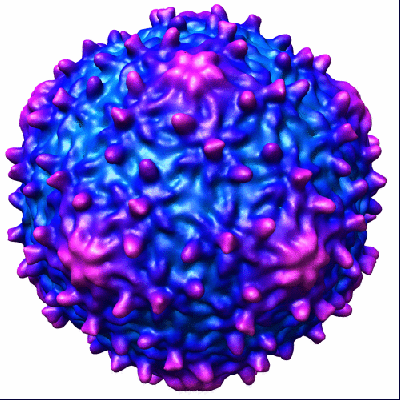
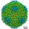
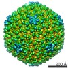
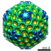
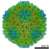
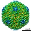
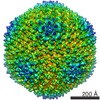
| Title | Genome sequence, structural proteins, and capsid organization of the cyanophage Syn5: a "horned" bacteriophage of marine synechococcus. | |||||||||
 Map data Map data | Bacteriophage Syn5 | |||||||||
 Sample Sample |
| |||||||||
| Biological species |  Synechococcus phage Syn5 (virus) Synechococcus phage Syn5 (virus) | |||||||||
| Method | single particle reconstruction / cryo EM / Resolution: 18.0 Å | |||||||||
 Authors Authors | Pope WH / Weigele PR / Chang J / Pedulla ML / Ford ME / Houtz JM / Jiang W / Chiu W / Hatfull GF / Hendrix RW / King J | |||||||||
 Citation Citation | Journal: J Mol Biol / Year: 2007 Title: Genome sequence, structural proteins, and capsid organization of the cyanophage Syn5: a "horned" bacteriophage of marine synechococcus. Authors: Welkin H Pope / Peter R Weigele / Juan Chang / Marisa L Pedulla / Michael E Ford / Jennifer M Houtz / Wen Jiang / Wah Chiu / Graham F Hatfull / Roger W Hendrix / Jonathan King /  Abstract: Marine Synechococcus spp and marine Prochlorococcus spp are numerically dominant photoautotrophs in the open oceans and contributors to the global carbon cycle. Syn5 is a short-tailed cyanophage ...Marine Synechococcus spp and marine Prochlorococcus spp are numerically dominant photoautotrophs in the open oceans and contributors to the global carbon cycle. Syn5 is a short-tailed cyanophage isolated from the Sargasso Sea on Synechococcus strain WH8109. Syn5 has been grown in WH8109 to high titer in the laboratory and purified and concentrated retaining infectivity. Genome sequencing and annotation of Syn5 revealed that the linear genome is 46,214 bp with a 237 bp terminal direct repeat. Sixty-one open reading frames (ORFs) were identified. Based on genomic organization and sequence similarity to known protein sequences within GenBank, Syn5 shares features with T7-like phages. The presence of a putative integrase suggests access to a temperate life cycle. Assignment of 11 ORFs to structural proteins found within the phage virion was confirmed by mass-spectrometry and N-terminal sequencing. Eight of these identified structural proteins exhibited amino acid sequence similarity to enteric phage proteins. The remaining three virion proteins did not resemble any known phage sequences in GenBank as of August 2006. Cryo-electron micrographs of purified Syn5 virions revealed that the capsid has a single "horn", a novel fibrous structure protruding from the opposing end of the capsid from the tail of the virion. The tail appendage displayed an apparent 3-fold rather than 6-fold symmetry. An 18 A resolution icosahedral reconstruction of the capsid revealed a T=7 lattice, but with an unusual pattern of surface knobs. This phage/host system should allow detailed investigation of the physiology and biochemistry of phage propagation in marine photosynthetic bacteria. | |||||||||
| History |
|
- Structure visualization
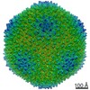
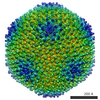
| Movie |
 Movie viewer Movie viewer |
|---|---|
| Structure viewer | EM map: SurfViewMolmilJmol/JSmol |
| Supplemental images |
- Downloads & links
-EMDB archive
| Map data | emd_1339.map.gz | 5 MB | EMDB map data format | |
|---|---|---|---|---|
| Header (meta data) | emd-1339-v30.xmlemd-1339.xml | 8.6 KB 8.6 KB | Display Display | EMDB header |
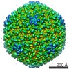
| Images |  1339.gif 1339.gif | 95.7 KB | ||
| Archive directory |  http://ftp.pdbj.org/pub/emdb/structures/EMD-1339ftp://ftp.pdbj.org/pub/emdb/structures/EMD-1339 http://ftp.pdbj.org/pub/emdb/structures/EMD-1339ftp://ftp.pdbj.org/pub/emdb/structures/EMD-1339 | HTTPS FTP |
-Validation report
| Summary document | emd_1339_validation.pdf.gz | 259.3 KB | Display | EMDB validaton report |
|---|---|---|---|---|
| Full document | emd_1339_full_validation.pdf.gz | 258.5 KB | Display | |
| Data in XML | emd_1339_validation.xml.gz | 5.9 KB | Display | |
| Arichive directory | https://ftp.pdbj.org/pub/emdb/validation_reports/EMD-1339ftp://ftp.pdbj.org/pub/emdb/validation_reports/EMD-1339 | HTTPS FTP |
-Related structure data
| Similar structure data |
|---|

-Links
| EMDB pages | EMDB (EBI/PDBe) / EMDataResource |
|---|
-Map
| File | Download / File: emd_1339.map.gz / Format: CCP4 / Size: 11.1 MB / Type: IMAGE STORED AS FLOATING POINT NUMBER (4 BYTES) | ||||||||||||||||||||||||||||||||||||||||||||||||||||||||||||||||||||
|---|---|---|---|---|---|---|---|---|---|---|---|---|---|---|---|---|---|---|---|---|---|---|---|---|---|---|---|---|---|---|---|---|---|---|---|---|---|---|---|---|---|---|---|---|---|---|---|---|---|---|---|---|---|---|---|---|---|---|---|---|---|---|---|---|---|---|---|---|---|
| Annotation | Bacteriophage Syn5 | ||||||||||||||||||||||||||||||||||||||||||||||||||||||||||||||||||||
| Voxel size | X=Y=Z: 5.43 Å | ||||||||||||||||||||||||||||||||||||||||||||||||||||||||||||||||||||
| Density |
| ||||||||||||||||||||||||||||||||||||||||||||||||||||||||||||||||||||
| Symmetry | Space group: 1 | ||||||||||||||||||||||||||||||||||||||||||||||||||||||||||||||||||||
| Details | EMDB XML:
CCP4 map header:
| ||||||||||||||||||||||||||||||||||||||||||||||||||||||||||||||||||||
-Supplemental data
- Sample components
-Entire : Syn5
| Entire | Name: Syn5 |
|---|---|
| Components |
|
-Supramolecule #1000: Syn5
| Supramolecule | Name: Syn5 / type: sample / ID: 1000 / Number unique components: 1 |
|---|
-Supramolecule #1: Synechococcus phage Syn5
| Supramolecule | Name: Synechococcus phage Syn5 / type: virus / ID: 1 / Name.synonym: Cyanophage Syn5 / NCBI-ID: 438482 / Sci species name: Synechococcus phage Syn5 / Virus type: VIRION / Virus isolate: STRAIN / Virus enveloped: No / Virus empty: No / Syn species name: Cyanophage Syn5 |
|---|---|
| Host (natural) | Organism:  Synechococcus (bacteria) / synonym: BACTERIA(EUBACTERIA) Synechococcus (bacteria) / synonym: BACTERIA(EUBACTERIA) |
| Virus shell | Shell ID: 1 / Name: capsid / Diameter: 600 Å / T number (triangulation number): 7 |
-Experimental details
-Structure determination
| Method | cryo EM |
|---|---|
 Processing Processing | single particle reconstruction |
| Aggregation state | particle |
-Sample preparation
| Buffer | pH: 8 / Details: 100mM NaCl, 100mM MgCl2, 50mM Tris |
|---|---|
| Grid | Details: 400 mesh copper grid |
| Vitrification | Cryogen name: ETHANE / Chamber humidity: 100 % / Chamber temperature: 298 K / Instrument: OTHER / Details: Vitrification instrument: Vitrobot |
- Electron microscopy
| Microscope | JEOL 2010F |
|---|---|
| Temperature | Average: 100 K |
| Details | 55360 magnification on CCD |
| Image recording | Category: CCD / Film or detector model: GATAN ULTRASCAN 4000 (4k x 4k) / Average electron dose: 10 e/Å2 |
| Electron beam | Acceleration voltage: 200 kV / Electron source:  FIELD EMISSION GUN FIELD EMISSION GUN |
| Electron optics | Illumination mode: FLOOD BEAM / Imaging mode: BRIGHT FIELD / Nominal defocus max: 5.0 µm / Nominal defocus min: 1.0 µm / Nominal magnification: 40000 |
| Sample stage | Specimen holder: Side entry liquid nitrogen-cooled cryo specimen holder Specimen holder model: GATAN LIQUID NITROGEN |
-Image processing
| CTF correction | Details: each particle |
|---|---|
| Final reconstruction | Applied symmetry - Point group: I (icosahedral) / Algorithm: OTHER / Resolution.type: BY AUTHOR / Resolution: 18.0 Å / Resolution method: FSC 0.5 CUT-OFF / Software - Name: SAVR / Number images used: 2616 |
